Biomedical Engineering Reference
In-Depth Information
thenaddedtothecorrespondingwells,andthemediawerechanged
every other day. PGA/PLA scaffolds alone (without cells) were cul-
turedininductivemediaasanothercontrol.After
in vitro
culturefor
oneweek,the constructswereimplanted
in vivo
to repair theosteo-
chondral defect.
Gross view and histology also demonstrated that the articu-
lar cartilage defect was fully repaired by engineered hyaline carti-
lage. Interestingly, an across section at the repair site showed that
the underlying cancellous bone defect was repaired by bone tissue
(Fig. 34.8) rather than the hyaline cartilage as shown in the previ-
ous study in which chondrocytes were used, indicating that BMSCs
candifferentiateintodifferentcelltypes
in vivo
,possiblyinducedby
different local environmental factors. Thus BMSCs may serve as a
better cell source for repairing articular cartilage defect if it is asso-
ciated with adefect of underlying bonetissue.
16
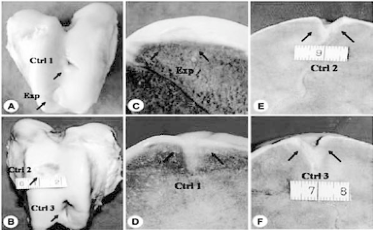
Figure 34.8.
Gross and cross-sectional view of repaired defects at 6
months postrepair. Arrows indicate the repaired regions. The experimental
defect exhibits a relatively regular surface (Exp, a), and the osteochondral
defectiscompletelyrepairedwithbothengineeredcartilageandbonewhen
observed at the cross section (c). The repaired surface of control group 1
remainsirregular(Ctrl1,a),buttheosteochondraldefectismostlyrepaired
at the cross section (d).The defectsin control group 2 (Ctrl 2, b, e) andcon-
trol group 3 (Ctrl 3, b, f) remain largely unrepaired at both cartilage and
bony layers. (Reprinted by permission from Ref. 16).


Search WWH ::

Custom Search