Biomedical Engineering Reference
In-Depth Information
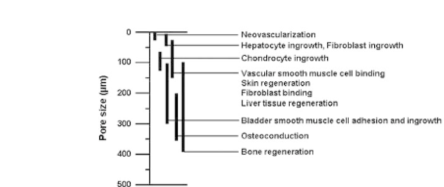
Figure 30.1.
Suggested optimum pore size ranges for different cells or
tissues (Ref. 22).
bladdersmoothmusclecelladhesionandingrowth,
17
100
∼
400
μ
m
forboneregeneration,
18
-
21
and200
mforosteoconduction
8
(summarizedinFig.30.1),dependingoftheporosityaswellasscaf-
fold materials used. Nevertheless of these investigations, more sys-
tematicevaluationonoptimumporesizerangesofscaffoldhasbeen
stillrequiredbecauseoftheabsenceofwell-definedscaffoldswitha
wide range of pore sizes. Thus, if the scaffold with a pore size gradi-
ent can be prepared, it will become a powerful tool for basic stud-
ies of the interactions between cells or tissues and scaffolds with
different pore sizes since the effect of pore size can be effectively
examined using one scaffold. Several techniques, including partic-
ulate leaching, phase separation, freeze-drying, gas foaming, fiber
bonding, and 3D printing have been used to fabricate porous poly-
mer scaffolds having a 3D pore structure.
1
Among them, the par-
ticulate leaching and freeze-drying methods, which can control the
pore size of scaffolds by porogen size and freezing temperature,
respectively,havebeencommonlychosentoevaluateoftheporesize
effect.
18
,
23
However, it is not possible from those techniques to fab-
ricate scaffolds with a pore size gradient. Also it is troublesome that
the scaffolds with different pore sizes are separately fabricated, and
thus low reproducibility of pore size distribution is still a limitation.
Recently,3Dprinting
24
andspinning
25
techniqueshavebeentriedto
fabricate scaffolds witha pore sizegradient.
∼
350
μ


Search WWH ::

Custom Search