Biomedical Engineering Reference
In-Depth Information
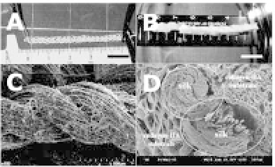
Figure 28.2.
Gross appearance (a, b) and scanning electron microscopy
(c, d) of the silk scaffold (a, c) and reinforced collagen-hyaluronan scaffold
with silk thread (b, d). (Circles: Cross-section of silk thread). Scale bar
=
a,
b: 1,000
μ
m; c, d
100. (Seo
et al
.
40
)
=
500
μ
m. Magnification
=
c, d
×
and physical properties,
38
but use of the silk material alone did not
enablesu
cient attachment orgrowth ofcells.
Recently, a reinforced composite silk scaffold was designed and
found to have the mechanical properties of a silk material, while
enablingincreasedadhesionandproliferationofcellsbylyophilized
collagen-hydroxyapatite (HA) substrates. The collagen-hyaluronan
substratesinthereinforcedsilkscaffoldalsoledtoincreasedratesof
cell migration when compared with silk. Moreover, these substrates
induced angiogenesis, which is essential for the initial phase of
repair of a damaged ligament (Figs. 28.2 and 28.3).
39
,
40
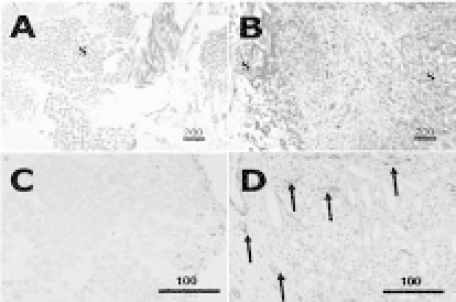
Figure 28.3.
Histological comparison of the silk scaffold (a, c) and com-
posite silk scaffold (b, d). The cross section was performed in the center of
the silk and composite scaffolds. (a, b: s
=
silk fiber; B: Blue color
=
new
synthesis collagen; H: arrow: blood vessels). Masson Trichrome stain: (a, b)
=×
40,scalebar
=
200
μ
m.CD31stain:(c,d)
=×
200,scalebar
=
100
μ
m.
(Seo
et al
.
40
) See also Color Insert.


Search WWH ::

Custom Search