Biomedical Engineering Reference
In-Depth Information
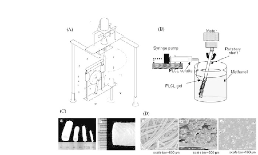
Figure 27.3.
Device (A) for gel-spinning molding technique (B) and
appearance(C)oftubularfibrousPLCLscaffoldspreparedbythetechnique.
SEM images of a double-layered PLCL scaffold (D, a: outer surface, b: cross-
section, and c: luminal surface).
blood vessels, such as the aorta, rather than for use in
in vitro
culture for tissue reconstruction.
22
FibrousPLCLscaffoldswere
enforced with a microporous inner layer for blocking of blood
leakage during implantation due to the large pore space on
the interior of nonreinforced PLCL scaffolds. Cylindrical shafts
were dip-coated with a PLCL/salt mixture and coupled to a gel-
spinning molding device for the formation of the outer fibrous
structure. The morphology of double-layered scaffold is shown
in (Fig. 27.3D). The individual fibers of the outer layer were
found to be crossed and/or fused together with neighboring
fibers (Fig. 27.3D-b). These cross-linked fibrous networks cause
the spun PLCL scaffolds to be open-pore structures and well-
interconnected between pores (Fig. 27.3D-a). An inner layer (Fig.
27.3D-c), which was a membrane with separated pores, was shown
to be well fused with the outer fibrous networks (Fig. 27.3D-b).
The scaffolds exhibited 550-670% elongation-at-break. The elas-
tic features of the scaffolds are shown in (Fig. 27.4A). The scaf-
folds exhibited a complete rubber-like elasticity being recovered


Search WWH ::

Custom Search