Biomedical Engineering Reference
In-Depth Information
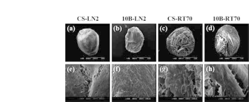
Figure 23.7.
SEM micrographs of the surface of the chitosan bead scaf-
folds cultured on chondrocytes after 14 d of culture. (a-d) Entire mor-
phology (magnification
×
40) and (e-h) magnified images (magnification
×
400).
7
proliferation of chondrocytes was exhibited only on the surface of
the CS-LN2 and 10B-LN2 bead scaffolds having small-size macrop-
ores but exhibited on both the surface and in the center of the CS-
RT70 and10B-RT70 bead scaffolds havinglarge-size macropores.
7
23.5 Chitosan Hydrogels
Biodegradable thermo-gelling hydrogels that undergo a sol-gel
transition with increasing temperature were studied by many
researchersintissueengineering.
30
Theadvantagesoftheinjectable
hydrogels are that they can fill any shape of a defect, may incorpo-
rate drugs and growth factors by simple mixing, and do not require
invasive surgery.
31
A block copolymer of poly-(ethylene oxide-
propylene oxide-ethylene oxide) (Pluronic, known as Poloxamer)
32
and the chitosan/
β
-glycerol phosphate (
β
-GP) system are well
known as thermogels.
33
Hoemann
et al
. reported that the thermo-
gelling hydrogels consisting of chitosan and
β
-GP and can support
in vitro
and
in vivo
accumulation of cartilage matrix by primary
chondrocytes, while persisting in osteochondral defects at least
one week
in vivo
.
34
Kim
et al
. also reported that the bone for-
mation from rat muscle-derived stem cells (rMDSCs) using an
injectable
in situ
-formingchitosangel
in vivo
wasexamined,andthe
rMDSCs survived well on the hydrogel created by the
in vitro
and
in


Search WWH ::

Custom Search