Biomedical Engineering Reference
In-Depth Information
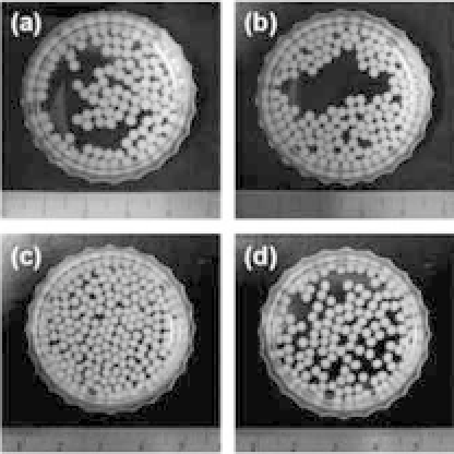
Figure 23.4.
Photographs of chitosan bead scaffolds. (a) CS-LN2,
(b)10B-LN2,(c) CS-RT70,and (d)10B-RT70.
7
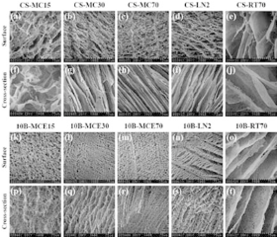
Figure 23.5.
SEMmicrographsofchitosanbeadscaffoldspreparedatvar-
ious temperatures and in various cooling media. (a-e, k-o) Surface and (f-j,
p-t)crosssectionofchitosanbeadscaffolds(magnification
×
400).Thedes-
ignation of each sample islisted in Table 23.2.
7


Search WWH ::

Custom Search