Biomedical Engineering Reference
In-Depth Information
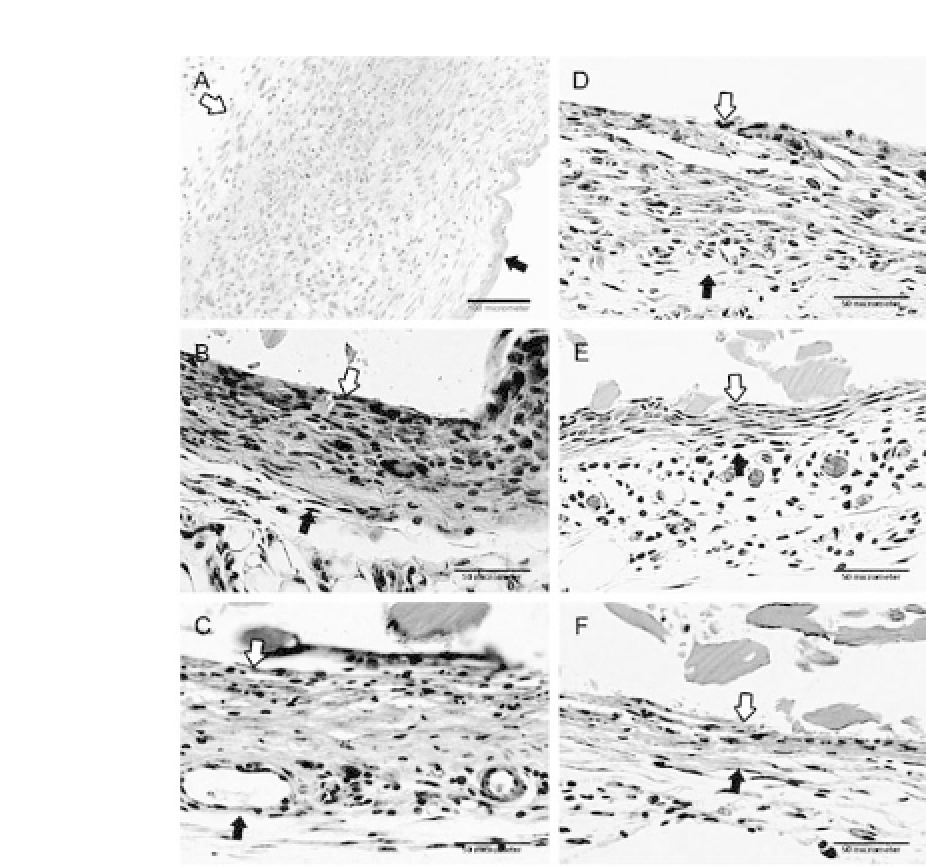
Figure 16.14.
PhotomicrographsofH&Estainsectionsofthedirectlybor-
dering tissue after PLGA, DBP hybrid PLGA films, and DBP. (a) The tis-
sue implanted with PLGA, bar length
=
100
μ
m (x100); (b-f) the tissue
implanted with 10% DBP/PLGA, 20% DBP/PLGA, 40% DBP/PLGA, 80%
DBP/PLGA, and DBP, respectively, bar length
=
50
μ
m (x400). Note that
the number of inflammatory cells and fibrous band thickness in vicinity to
the tissue-implanted samples decreased as the DBP content in PLGA films
increased.Polymer-tissueinterfacesurfacesareindicatedbyawhitearrow.
The fibrous wall thickness is represented by black and white arrows. See
also ColorInsert.
cytokines such as IL-1
β
.TNF-
α
and IL-1
β
are potent stimula-
tors of fibroblast growth.
25
Therefore, temporospatial expression
of these pro-inflammatory mediators allows fine-tuning of the
inflammatoryresponse.
6
DBPshavebeenreportedasbiocompatible


Search WWH ::

Custom Search