Biomedical Engineering Reference
In-Depth Information
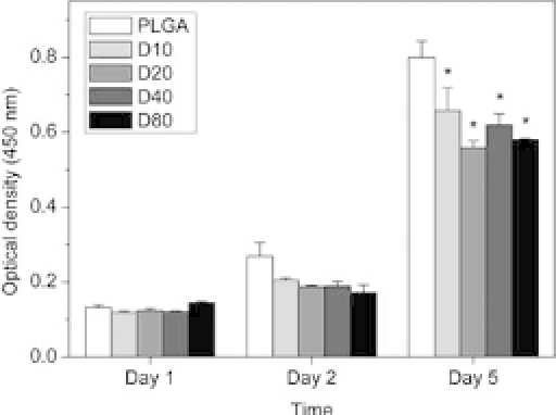
Figure 16.8.
The number of viable HL-60 cells on PLGA and DBP/PLGA
scaffoldsatDays1,2,and5,asdetermined bytheMTTcolorimetric assay. *
corresponds to
p
<
0.05 in comparison with PLGA scaffolds for each day.
and the 10% DBP/PLGA scaffold at Days 2 and 3. Next we evaluated
the viability of HL-60 cells on samples and did not find statistical
significances between PLGA and DBP hybrid scaffolds at Days 1 and
2.BycultureDay5,proliferationofHL-60cellswithPLGAincreased
slightly more than withDBP/PLGA scaffolds(Fig. 16.8).
16.3.2
Inflammatory Cytokine Expression
Toelucidatethecellularresponsesassociatedwithinflammationon
sample films, we measured the level of mRNA expression of tumor
necrosis factor-
α
(TNF-
α
) and IL-1
β
from HL-60 cell in 48 hours
after culture with PLGA or DBP/PLGA films, as shown in Fig. 16.9.
TNF-
α
mRNA in HL-60 highly expressed following PLGA films com-
pared with DBP/PLGA films; it was significantly lower following
DBP/PLGA
films than PLGA films, with increases in contents of DBPs: 10%,
20%,40%,and80%ofDBPs(
p
<
0
.
005,
p
<
0
.
0001,
p
<
0
.
00005,
and
p
<
0
.
00005, respectively) (Fig. 16.10). The intensity of TNF-
α
expression of PLGA films was significantly 10 times higher or more
thanthatof40%DBP/PLGAfilms.HL-60cellswith80%DBP/PLGA
films rarely expressed TNF-
mRNA
expressiondecreasedmarkedlywith40%and80%DBP/PLGAfilms
α
mRNA. Similarly, IL-1
β


Search WWH ::

Custom Search