Biomedical Engineering Reference
In-Depth Information
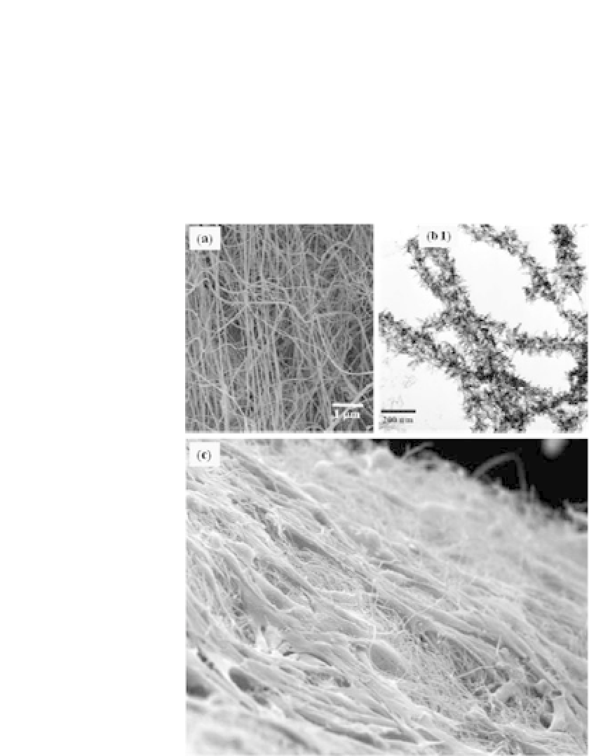
to hundreds of nanometers as the sol concentration was varied
(Fig. 14.2). When rBMSCs were cultured on the bioactive glass
nanofiber, they favorably migrated over and secreted an osteoblas-
tic differentiation phenotype. The differentiation level was signifi-
cantlyhigheronthenanofiberformofbioactiveglassthaneitheron
the dense sintered bioactive glass or on the nanofiber form of the
Figure 14.2.
Bioactive inorganic nanofibers obtained by electrospinning:
(a) SEM image of bioactive glass nanofibers produced by electrospin-
ning and heat treatment; (b) TEM image of acellular bone bioactivity of
nanofibers, showing the formation of bone mineral-like apatite on the
surface after soaking in SBF for 3 days (adapted with permission from
Ref. 25; copyright 2006 Wiley-VCH Verlag GmbH & Co.); (c) Rat BMSCs
grown on bioactive glass nanofibers for 7 days showing cells were viable
with active cytoplasmic extensions in contact with the underlying nanofi-
brous substrate.


Search WWH ::

Custom Search