Biomedical Engineering Reference
In-Depth Information
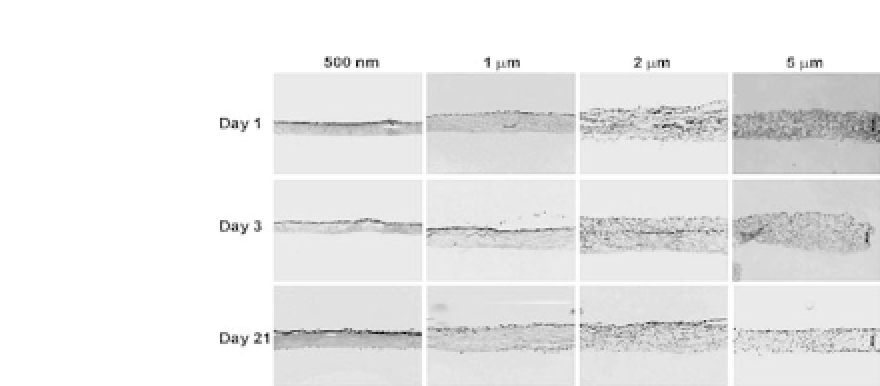
Figure 10.6.
Cellular infiltration into the electrospun scaffolds with var-
ious fiber diameters. NIH/3T3 fibroblasts were seeded into electrospun
PCL/collagen scaffolds up to 21 days. Nuclei stained by DAPI (
×
40 magnifi-
cation) (unpublished data).
related to the pore area in electrospun scaffolds. Overall, obtain-
ing adequate cellular infiltration into electrospun nanofibrous scaf-
folds while maintaining their mechanical properties and structural
integrity is stilla major challenge.
10.3.4
Cellular Differentiation
Cellular differentiation on electrospun fibers is closely related to
cell-substrate interactions. Neural stem cell (NSC) proliferation can
bepromotedusingfibroblastgrowthfactor-2(FGF-2).
39
Inaddition,
NSC can be preferentially differentiated into neurons using retinoic
acid and forskolin,
40
into astrocytes using leukemia inhibitor fac-
tor (LIF) and bone morphogenic protein (BMP),
41
and into oligo-
dendrocyteswithinsulin-likegrowthfactor(IGF)orplatelet-derived
growth factor (PDGF).
42
,
43
However, the effect of topographical
cues on NSC proliferation and differentiation is poorly under-
stood. It remains unclear how topographical features (specifically,
nanofiber diameter and alignment) influence stem cell proliferation
and differentiation, and this is partially due to a lack of reliable
methods for producing fibers with well-defined diameters. System-
atic characterization of nanotopographical regulation of cell behav-
ior is important in understanding and eventually engineering on an


Search WWH ::

Custom Search