Biomedical Engineering Reference
In-Depth Information
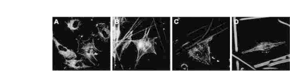
Figure 10.2.
Cytoskeletal and focal adhesion of human aortic endothelial
cells on electrospun nanofibers with various fiber diameters; (A) 500 nm,
(B)1
μ
m,(C)2
μ
m,and(D)5
μ
m(
×
600magnification).Red:F-actin,Green:
vinculin (unpublished data).
focal adhesion of endothelial cells (EC) on electrospun polycapro-
lactone (PCL)/collagen scaffold with different fiber diameters. EC
on nano-scaled fibers (0.5
μ
m, Fig. 10.2a) show a better-developed
cytoskeletal organization and improved focal adhesion compared
with other fiber diameters. Because the fiber diameters of electro-
spun nanofiber scaffolds are orders of magnitude smaller than the
size of most cells, the cells are able to organize around the fibers or
spread and attach to adsorbed proteins at multiple focal points.
14
,
15
Furthermore,Finne-Wistrand
et al
.haveshownthattheelectrospun
nanofibrous mats can enhance adhesion and proliferation of mes-
enchymal stem cells compared with a flat smooth surface.
16
Severalstudieshavedemonstratedthatelectrospunfibrousscaf-
folds can enhance cellular responses, including cell adhesion and
maintenanceofcellphenotypemaintenance.
17
-
20
Shih
et al
.demon-
strated the adhesion, proliferation, motility, and differentiation of
human mesenchymal stem cells on electrospun type I collagen
nanofibersofdifferentdiameters.
21
Theseindicatethatnano-scaled
fibers can support initial cell adhesion, which then affects further
cell proliferation and differentiation.
10.3.2
Cell Alignment
Nanofiber scaffolds are known to guide cellular attachment and ori-
entation.Assuch,nanofibersfabricatedbyelectrospinningprovides
a controlled environment that allows for enhanced cellular orien-
tation that leads to accelerated tissue function. Skeletal muscle is a
tissue that possesses unidirectional cellular orientation to function


Search WWH ::

Custom Search