Biomedical Engineering Reference
In-Depth Information
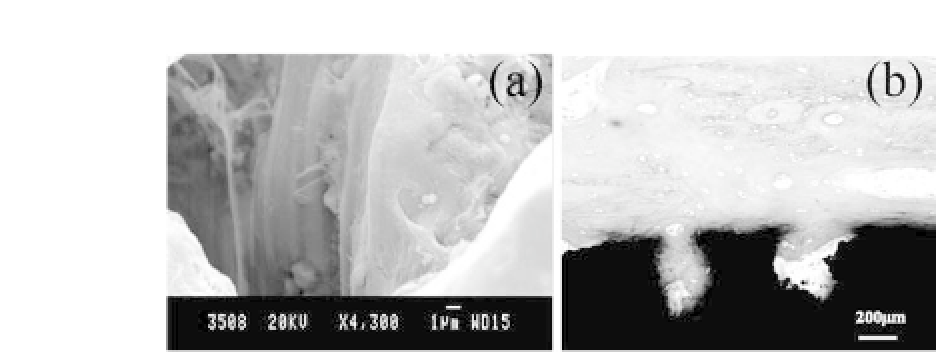
Figure 3.6.
In vitro
and
in vivo
tests of 3D macroporous Ti-based metal
scaffolds produced by CF-HIP with NH
4
HCO
3
as a space holder. (a) SEM
image of cell in-growth on internal exposed surface of the NiTi scaffold; (b)
Optical image of bone tissue (pink color) growth on the entire exposed sur-
face of the Ti scaffold (black color). See also Color Insert.
The porosity can be controlled from 21% to 56% (in volume),
and the open porosity can reach 70% using thismethod.
15
,
17
Our previous investigations show that both 3D porous NiTi and
TiscaffoldsproducedbyCF-HIPexhibitgoodmechanicalproperties
and similar Young's modulus with human bone and that cells can
attach smoothly and grow on the surface of the internal pores, as
illustratedinFig.3.6a.Afterashort-term(3months)
in vivo
test,the
porous scaffolds also bode well for bone tissue in-growth, as shown
in Fig.3.6b.
3.3 Natural Growth and Characterization
of 1D Nano Titanates
In view of the hierarchical organization of bones on the nano scale
and the fact that nanophase materials significantly influence tis-
sue acceptance and cell behavior
18
−
20
it is necessary to modify the
surface of 3D macroporous Ti-based metal scaffolds to achieve a
hierarchical structure on the nano scale to enhance its acceptabil-
ity by nano HA crystals, tropocollagen, and other proteins from
bone cells. Because of the 3D macroporous structure of Ti-based
metal scaffolds, it is very di
cult to treat the complex topographies
using traditional line-of-sight techniques such as laser nitriding,


Search WWH ::

Custom Search