Biomedical Engineering Reference
In-Depth Information
Õ
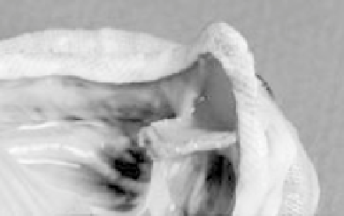
5.7 Photo of an explanted Edwards Lifesciences S.A.V.
valve, showing the
problems of excessive trimming of the aortic wall during production. The too-
thin wall tissue was unable to support the leaflets during use and they pulled
out from the surrounding stent. The solution to this problem was to provide
adequate wall tissue to provide the necessary support for the leaflets. (Photo
taken from Jamieson WRE, et al. (1999), `Carpentier-Edwards standard and
supra-annular porcine bioprostheses: comparison of technology,' Ann Thorac
Surg, 67, 10. Reprinted with permission from The Society of Thoracic
Surgeons. All Rights Reserved.)
· Are the leaflets inside or outside the stent? The Ionescu±Shiley valve wrapped
the tissue around the outside of the stent, giving the valve maximum orifice
area, while the PERIMOUNT
Õ
valve wrapped the tissue around the inside of
the stent. Ultimately, the Ionescu±Shiley valve failed due to poor long-term
durability. This is because the leaflets were being rubbed against the cloth-
covered stent during valve closure, which is a period of maximum pressure.
The PERIMOUNT
Õ
design resulted in leaflets occasionally contacting the
stent during valve opening, which is relatively low pressure. Tissue contact to
the stent during valve closure resulted in maximum abrasion and, thus,
significantly shorter durability for the Ionescu±Shiley valve. Figure 5.8
depicts the differences between these two valves and shows a close-up
photograph of a leaflet in close proximity of a cloth-covered stent. Recently,
St. Jude Medical has commercialized the Trifecta
TM
valve, where the leaflets
are wrapped outside of the stent, but the stent is covered with thin pericardium,
to try to minimize tissue abrasion. Long-term results will be required to
determine if this approach will be successful in preventing tissue abrasion.
· How are the leaflets attached to the stent, particularly at the commissure tips? A
critical design flaw of the Ionescu±Shiley valve was the use of an aligning suture
over the top of the leaflets over the stent. As shown in Fig. 5.9, this alignment
suture became the site of early failure, due to stress concentration around the
suture, causing the tissue to yield and ultimately tear. Later design iterations
removed this alignment suture and lowered the profile of the valve, but the valve
still failed clinically due to stent abrasion. In accelerated wear testing this
abrasion resulted in perforation of the leaflet all along the attachment line to the
stent, beginning at the top of the commissure, the site of maximum stress.