Biomedical Engineering Reference
In-Depth Information
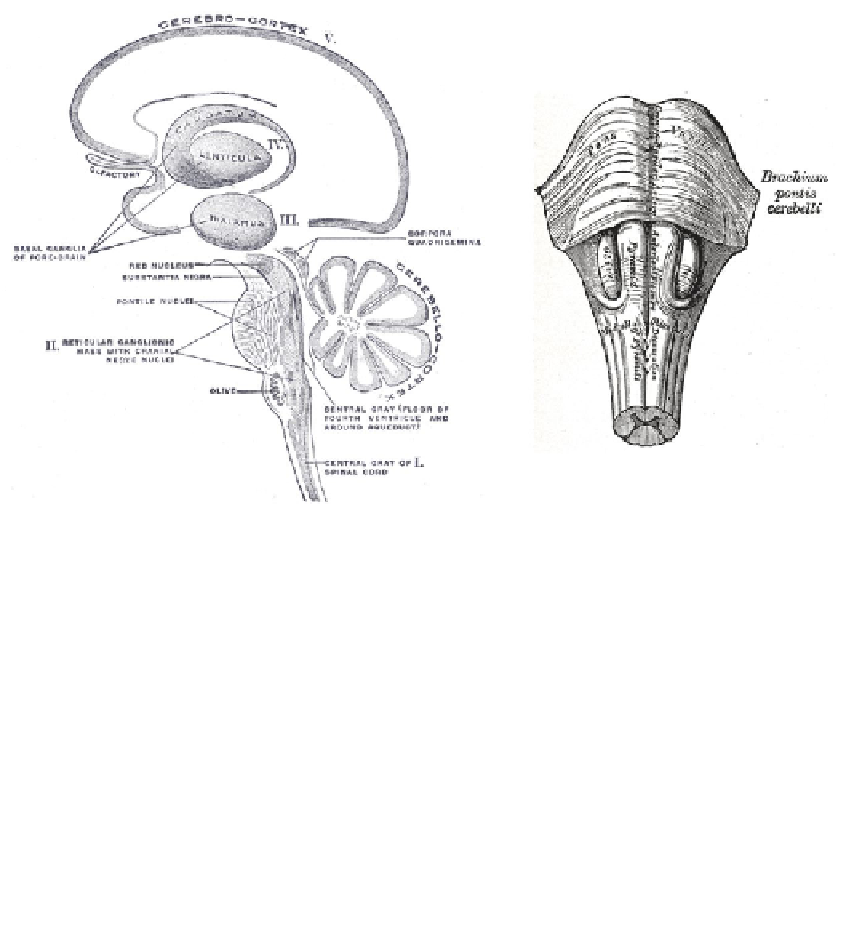
Figure 1.2:
Gross anatomy of the brain.
1.3.2 Brain Systems
The gross anatomy of the brain consists of both tissue (neurons and glia) as well as chambers called
ventricles that are filled with cerebral-spinal fluid. The tissue is organized into three regions based upon
embryonic development.
The
hindbrain
, or Rhombencephalon, is an offshoot of the spinal cord and was inherited from
the reptile brain. It is primarily involved in involuntary control of basic functions such as breathing and
heart rate. In addition to forming the lower portion of the brain stem, it also includes the cerebellum, a
relatively large outcropping at the base of the brain that controls motor movements and relays signals to
the spinal cord.
Table 1.3:
Myelencephalon
Medulla oblongata
Metencephalon
Contains 4th Ventricle
Pons
Relay sensory information
Cerebellum
Integration of sensory and motor
Reticular Formation
Rudimentary eye movements
The
midbrain
, or Mesencephalon, forms the upper part of the brain stem. Its primary function is
to connect the lower brain stem with higher-level brain structures. In size, it is much smaller than the
other two embryonic regions. It is involved in involuntary motor control and sensation.