Biomedical Engineering Reference
In-Depth Information
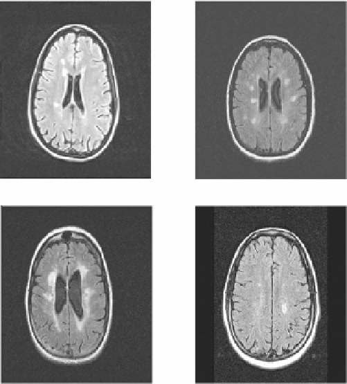
Figure 1.14: Sample slices from FLAIR MRI studies of patients with MS. Hy-
perintense regions in the brain are indicative of plaques caused by MS.
to clinically observed deficits [37]. As well, quantification of MRI studies of MS
have yet to be perfected, and most studies of MS do not generate enough MRI
data to evaluate the nuances in the application of MRI to study MS.
It is desirable to develop computer tools to assist experts in the study of MS
using MR imaging. Allowing a computer to automatically identify normal and
abnormal brain tissue would free an expert from the arduous task of manually
examining each slice of a study, while generally increasing the reproducibility of
the identification by removing the subjectivity of the human observer. Medical
registration tools would also be useful for automatic, retrospective alignment of
patient studies, taken at different points in time, to allow for qualitative compar-
ison of different studies of a patient of the course of his or her treatment. This
type of analysis would aid the expert in deciding if the disease is responding
well to the present treatment, or if a change in the treatment is warranted.
Typically in an MRI study, a patient is placed in the scanner with little regard
for positioning of the anatomy of interest. The only constraints are that the

