Biomedical Engineering Reference
In-Depth Information
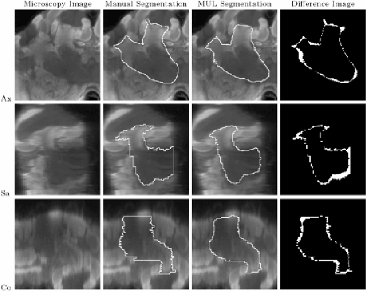
Figure 11.15: A typical segmented structure: right ventral mushroom body
(SI
=
0.86).
Columns from left to right
: microscopy image, contour from manual
segmentation, contour from automatic segmentation (MUL paradigm), and dif-
ference image between manual and automatic segmentation. The white pixels in
the difference image show where manual and automatic segmentation disagree.
Rows from top to bottom
: axial, sagittal, and coronal slices through the right
ventral mushroom body.
results of all strategies. Only slightly better results were achieved by selecting a
different individual atlas for each raw image, based on the NMI after non-rigid
registration criterion discussed in section 11.4.2. The AVG strategy, segmenta-
tion using an average shape atlas, outperformed both the IND and SIM strategies,
but was itself clearly outperformed by the MUL strategy. Our results therefore
show that the multiclassifier approach to atlas-based segmentation produced
substantially more accurate segmentations than the other three strategies. This
finding is, in fact, statistically significant when performing a t-test on the SI val-
ues for all structures over all segmentations, which confirms the experience of