Biomedical Engineering Reference
In-Depth Information
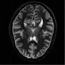
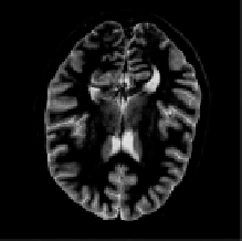
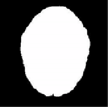
Figure 9.12: The original slice of anatomical MRI brain image ((top left), origi-
nal superimposed over the true deformation (top right), the recovered deforma-
tion versus the true deformation (bottom left), and the mask used to calculate
the warping index (bottom right). (Color Slide)
9.4.10.1
Registration of Artificially Deformed MRI Brain Slices
To illustrate the behavior of the algorithm, we show its performance when recov-
ering a known deformation of a 2D slice of an anatomical spin-echo MRI volume
of the brain.
9
We use here artificially-deformed images because the knowledge
of the ground truth permits us to better judge the performance of the algorithm.
The original image of size 256
×
256 pixels is shown in Fig. 9.12, top-left.
We use a cubic spline control grid with one knot for every 32 pixels. We warp
the image with a deformation belonging to the warp space and consisting of
displacements up to 15 pixels.
10
The warped image is superimposed on the
original in Fig. 9.12, top-right. Then the automatic registration algorithm is run
with the stopping threshold set to 0.5 pixels for all levels except the last, where
we set it to 0.1 pixels. The recovered deformation was used to warp again the
original image. Its warped version is shown superimposed on the image warped
9
First author's brain. Images courtesy of Arto Nirkko from Inselspital Hospital, Bern,
Switzerland.
10
Approximately 14 mm.