Biomedical Engineering Reference
In-Depth Information
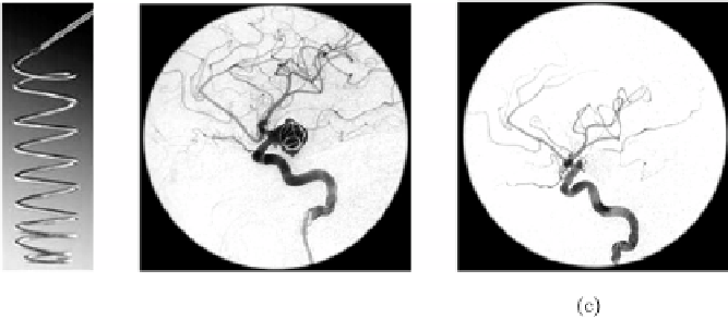
Figure 5.3:
Process of endovascular embolization with GDCs. (a) An example
of GDC. (b) Digital substraction angiography of an embolized aneurysm using
GDC coils. Introduction of the GDC inside the aneurysm by catheterization.
(c) Final result of the embolization. The placement of the coil promotes blood
clotting inside the aneurysm. The clot avoids blood flow inside the aneurysm
as is appreciated in the DSA image. The progressive reduction of intracranial
blood flow pressure avoids its rupture.
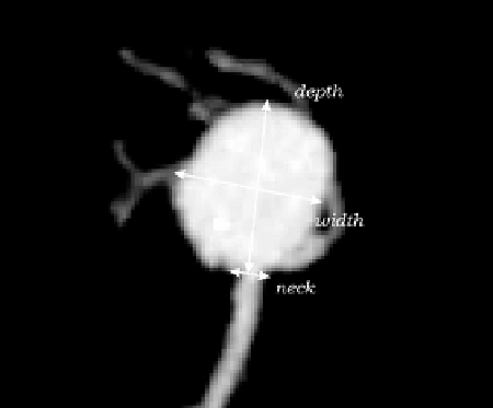
Figure 5.4: Maximum Intensity Projection (MIP) of a brain aneurysm recon-
structed from a CTA image. The measurements of the neck, dome width and
depth needed to carry out the selection of the coil size in the endovascular
treatment are indicated over the MIP.