Biology Reference
In-Depth Information
A
Signal initiation
H
H
H
E1
Dyn
Dyn
β
γ
β γ
α
P
GTP
H
P
GRK2
“Signalsome”
assembly
A
rr
E2
P
P
Early
endosome
Arr
G protein-dependent
signaling
E2
H
A
rr
E2
P
P
Arrestin-dependent
signaling
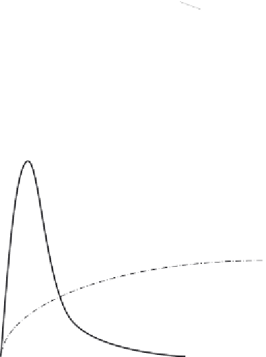
B
G protein-dependent signaling
Second messenger dependent
Arrestin-dependent signaling
Signalsome dependent
Seconds to minutes
Minutes to hours
Time range
Figure 5.2 Arrestin scaffolds impose spatial and temporal regulation of signaling path-
ways. (A) Upon agonist (H) binding, GPCRs engage heterotrimeric G proteins, activating
G protein-regulated effectors (E1) at the plasma membrane. Within seconds, GRK phos-
phorylation of the activated receptor creates high-affinity arrestin-binding sites. Arrestin
binding uncouples the receptor from heterotrimeric G proteins while targeting it for
endocytosis. As arrestins translocate to the receptor, they recruit additional catalytically
active proteins (E2) into receptor
-
arrestin signalsomes. These stoichiometric signaling
complexes transmit a distinct set of signals as the receptor internalizes and transits the
intracellular compartment. (B) G protein-dependent signaling is characterized by rapid
onset followed by waning intensity, reflecting desensitization due to receptor phos-
phorylation by second messenger-dependent protein kinases and GRKs, and arrestin
binding. In contrast, arrestin-mediated signals are of slower onset and often sustained
in duration. (A) Reproduced from Ref.
33
.


Search WWH ::

Custom Search