Biomedical Engineering Reference
In-Depth Information
implants that are extremely wear resistant can also be
made, and this is proving beneficial in the case of
some prosthetic hip designs.
Initially, the interest in CFR PEEK was largely
academic, but in the intervening years interest has
grown and the material is becoming a commercially
viable alternative to implant grade metallic alloys for
applications requiring high strength and fatigue
resistance. It is also being selected for the commer-
cial development of implantable load-bearing
medical devices, in which compatibility with modern
imaging technologies is a key concern, because
medical imaging is an important diagnostic tool and
it is desirable that the materials used in the
manufacturing of medical implants should be
compatible with a broad range of techniques.
In the case of X-rays, radiographically CFR PEEK
and natural unfilled PEEK polymers are transparent
(radiolucent), and this can be beneficial for a detailed
clinical inspection of the implant site. However, in
some cases it is necessary to have at least some level
of radiopacity, because the device may be required to
be observed using radiography for the medical team
to follow up on the clinical progress of the surgery.
Unfilled PEEK can be made moderately opaque by
the addition of a strongly X-ray absorbing filler at an
appropriate concentration, therefore called “image
contrast” grades. For this reason PEEK polymer has
been selected for the development of radiolucent
vertebral body replacement and other devices, which
enable the complete inspection of tissue and bone
growth via conventional imaging techniques.
Another benefit with medical devices that use
radiolucent materials is that they require less inten-
sive X-rays while retaining image accuracy and
definition; thus, overall patient exposure to radiation
is reduced.
Other medical imaging methods, such as computer
tomography (CT) and magnetic resonance imaging
(MRI), are not “metal friendly” and the presence of
metallic implants in the area being scanned can
significantly and negatively impact the quality of the
resulting images.
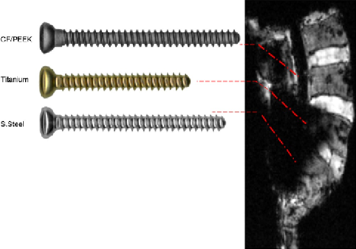
Figure 3.2
shows an example MRI
scan of a spine phantom with three implanted screws,
made from CFR PEEK, titanium, and stainless steel,
in upper, middle, and lower parts, respectively. It can
be seen that the image quality suffers significantly
from dark shadowing around the metallic implants,
whereas the image is significantly clearer adjacent to
the CFR PEEK screw.
In the case of CT images, metallic materials can
create artifacts where the signal from the directed
X-ray beam, required by the CT imager to reconstruct
the full
image, cannot penetrate the radiopaque
Figure 3.2
MRI image of CF/PEEK-OPTIMA, titanium, and stainless steel screws implanted in a phantom, illustrating
the dark shadowing that occurs around metallic implants. Photo courtesy of Icotec AG.