Biomedical Engineering Reference
In-Depth Information
plates (Carboxy: Orthodynamics Ltd., Dorset, UK)
have been used to treat over 1000 fractures since
1982, according to the literature from the manufac-
turer. PEEK fracture plates are currently available
through CarboFix Orthopedic Ltd. (Herzeliya,
Israel). CarboFix has a platform material made of
endless carbon fibers embedded in PEEK. Included
in their product portfolio, the Piccolo Nailing system
includes intramedullary nails used in the treatment of
humeral, tibial, femoral, and hip fractures. The
Piccolo Plating System provides plating options for
proximal humerus, distal radius, and long bone
diaphyseal fractures.
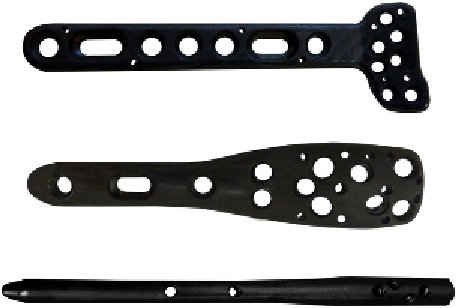
Figure 15.3
shows a few of the
PEEK composite devices currently marketed by
CarboFix.
Metallic locking plates and intramedulary nails
continue to dominate the field of internal fracture
fixation, as they have for over 100 years
[9,35,36]
.
Despite their utility, polymer implants still account
for only a small fraction of fracture fixation devices
that have been deployed clinically over the past two
decades. In the United States alone, over 600,000
internal fracture fixation procedures were performed
in 2006
[1]
. Although devices entirely comprised of
PEEK have not taken over the market, a number of
trauma devices are finding an ancillary role for
PEEK. Biotech International (Salon-de-Provence,
France) has added PEEK locking devices to bone
plates to increase stability with the system and
prevent the backing out of screws. The locking of
screws within the plate further allows fixed angle and
polyaxial screw positioning, functionality that mini-
mizes the need for plate
e
bone contact, long thought
to be associated with local bone absorption. This
concept has also been transferred to interlocking
screws in humeral nailing constructs. Yanez et al.
[37]
presented a new concept for osteosynthesis in
osteoporotic fractures that utilizes a PEEK locking
nut contralateral to the plate. The screw locking
element uses a pre-drilled tunnel in the center of the
nut that is threaded by the advancing screw. PEEK
inlays have also been used to prevent backing out of
screws used with the Targon-PH humeral nail
[38]
.
To the authors' knowledge, there have been no
clinical reports of the performance of PEEK mate-
rials as implanted fracture fixation devices in human
patients. More information about spinal fusion
applications for PEEK and its composites can be
found in Chapter 13.
15.3 Principles of Arthroscopic
Repair
The development of arthroscopic techniques has
revolutionized the repair of tendons, ligaments, and
other soft tissues. Arthroscopy minimizes the entry
port and uses an endoscope, or a small camera, along
with a set of small instruments to provide treatments
within a joint. These instruments are implemented
through a set of small, approximately 1 cm, incisions.
Prior to the use of such techniques, all soft tissue
repairs were done via either an open or mini-open
exposure. The advantage of the arthroscopic tech-
nique includes a significant decrease in pain as well
and surgical site problems that often exist far after the
repaired ligaments and tendons have healed.
Although the majority of arthroscopic procedures are
to repair torn or damaged menisci and anterior
cruciate ligaments (ACLs) in the knee, suture anchor
devices used in the shoulder have seen the most
dynamic
recent
development
in
terms
of
biomaterials.
Bone suture anchors to fasten ligaments, tendons,
and capsules to bone have helped shift open surgeries
toward arthroscopic techniques. Anchors are
embedded into the bone and serve to tie down soft
tissue to bone via a suture passing through its eyelet.
Some of the most common procedures that utilize
suture anchors are rotator cuff repairs, Bankart
repairs (reattachment and tightening of torn labrum
and ligaments to the shoulder), and superior labral
anterior posterior (SLAP) repairs. The anchors'
service is temporary as they are only required to stay
Figure 15.3
Piccolo PEEK composite devices by Car-
boFix, from top to bottom: Distal Volar Radius Plate,
Proximal Humeral Plate, and Proximal Humeral Nail.
The image is reprinted here with the permission of the
manufacturer.