Biomedical Engineering Reference
In-Depth Information
confer some subtle influence on cells
[31]
, so the
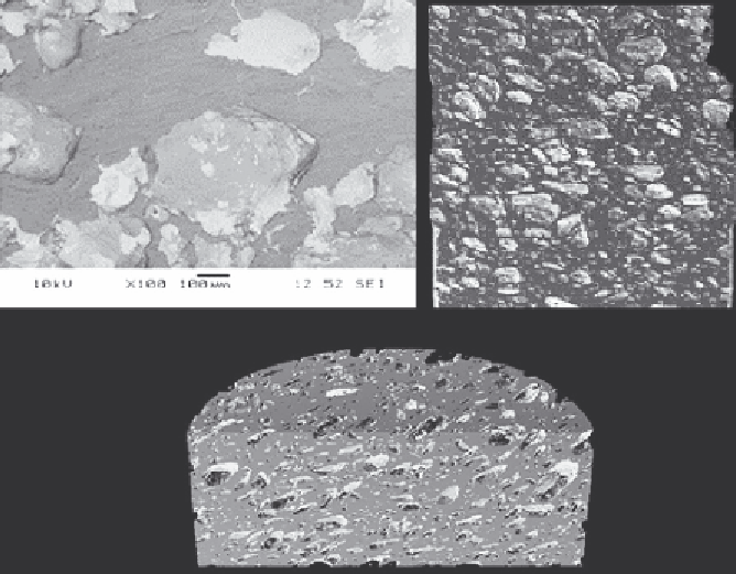
contrasting topography observed between porous and
machined samples may also influence the behavior of
surrounding tissue. When assessed using scanning
electron microscopy (SEM) and computed tomog-
raphy (CT), the porous PEEK had a porosity of
approximately 50% and a mean pore size of 70
m
m
(
Fig. 12.3
). A 1 cm path of the surface was measured
using a surface roughness analyzer (Federal Prod-
ucts) at three locations parallel to the axis of the
implant. This confirmed that the porous PEEK
surface presented at the implant
e
bone interface is
rougher (
R
a
for intervertebral body fusion devices
. The results
showed an overall permanent deformation of around
25
m
m in compression after 5 million cycles over
6 days. Parameters were as follows: 10 Hz;
F
max
:
2.0 kN, corresponding to 10 N/mm
2
;
F
min
: 0.2 kN,
giving a load ratio of 10. Samples were 13
15
195 mm
2
.
4 mm “cervical” squares
¼
12.5.3 In Vitro Testing
Cell culture on this version of a porous PEEK-
OPTIMA has been previously reported by Tandy
et al.
[32]
. Cultures of marrow stromal cells were
seeded onto porous PEEK samples (7.25 mm in
width, 13.5 mm in length, and 4.25 mm in thickness).
After varying time points (7, 14, and 21 days), the
cultures were assessed for total DNA, alkaline
phosphatase, osteopontin, and calcium to indicate
stages of proliferation, differentiation and minerali-
zation, respectively. Researchers at the University of
Oklahoma observed that the production of extracel-
lular mineralized matrix by the cells on the porous
samples occurred within the study duration (21 days)
and was detected by calcium deposition assay.
Energy dispersive X-ray spectroscopy (EDS) anal-
ysis of the end study duration samples detected levels
of carbon, phosphorus, and calcium. The morphology
of the cells under SEM showed a polygonal cell
shape, which is
¼
6.5
m
m) than machined PEEK samples
(
R
a
¼
3.5
m
m).
12.5.2 Mechanical Testing
Mechanical properties of the porous material were
measured according to ISO test procedures (
n ¼
5)
and showed the following mean properties: ISO
527
d
tensile strength (porous 14.5 MPa, PEEK
105.5 MPa), ISO 180
d
strain at break (porous 3.5%,
PEEK 49.5%), impact strength (porous 3.6 kJ/m
2
,
PEEK 7.7 kJ/m
2
), ISO 178
d
flexural strength
(porous 21.6 MPa, PEEK 174.6 MPa), and flex
modulus (porous 0.8 GPa, PEEK 4.4 GPa).
Testing that was specifically relevant to a spinal
device was also performed, ASTMD695/ISO604 and
Dynamic fatigue test ASTM F2077-03
dTest method
typical of
the differentiated
Figure 12.3
Porosity of the porous
PEEK samples was evaluated
using SEM and micro-CT.
(a)
(b)
(c)