Biomedical Engineering Reference
In-Depth Information
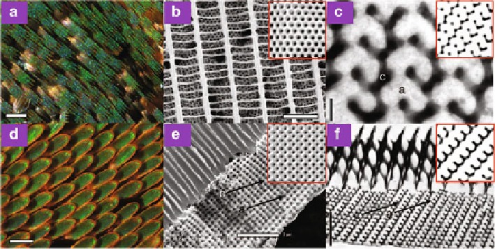
Fig. 8.33
Nanostructures for structural coloration in the scales of lycaenid and papilionid
butterflies. (
a
) Optical microscopic image of the ventral wing cover scales of
C. gryneus
.(
b
)SEM
image of the dorsal surface of a
C. gryneus
scale showing disjoint crystallites beneath windows
created by a network of parallel, longitudinal ridges and slender, spaced cross-ribs. The inset shows
the simulated SEM (111) projection from a thick slab of a level set single gyroid nanostructure. (
c
)
TEM image of the
C. gryneus
nanostructure showing a distinctive motif, uniquely characteristic
of the (310) plane of the gyroid morphology. The inset shows a matching simulated (310) TEM
section of a level set single gyroid model. (
d
) Optical microscopic image of the dorsal wing
cover scales of
P. sesostris
.(
e
) SEM image of the lateral surface of the wing scale nanostructure
of
P. sesostris
showing fused polycrystalline domains beneath columnar windows created by a
network of ridges and spaced cross-ribs. The fractured face features a square lattice of air holes in
chitin. The inset shows the simulated SEM (100) projection from a thick slab of a level set single
gyroid nanostructure. (
f
) TEM image of the
P. sesostris
nanostructure showing a distinctive motif,
uniquely characteristic of the (211) plane of the gyroid morphology. The inset shows a matching
simulated (211) TEM section of a level set single gyroid model. The labels
c
and
a
in (
c
), (
e
), and
(
f
) indicate chitin and air void, respectively. Scale bars: (
a
)and(
d
) 100
m; (
b
)2.5
m; (
c
) 200
nm; and (
e
)and(
f
)2
m (Reproduced from [
137
])
In additional to multilayers, 3D photonic crystals are also exploited in butterflies
for structural coloration, e.g., in papilionid and lycaenid butterfly scales [
129
-
137
].
Revealed 3D photonic structures exhibit various forms and complexity, attributed to
simple cubic [
129
] and face-centered cubic [
130
] structures, and gyroid structures
[
135
-
137
].
The wings of the butterflies
Callophrys gryneus
(Lycaenidae) and
Pa rid es
sesostris
(Papilionidae) display vivid structural colors. Structural characterizations
based on electron microscopy and small angle X-ray scattering [
137
] show that
these wing colors are caused by a gyroid (
I
4
132) photonic crystal in the scales, a
bicontinuous triply periodic structure of the network of chitin and air, as shown in
Fig.
8.33
. The measured lattice parameter is about 306 nm for
C. gryneus
and about
288 nm for
P. sesostris
. The estimated filling fraction is about 0.34 and 0.3 for
C.
gryneus
and
P. sesostris
, respectively. Photonic band structure calculations revealed