Biomedical Engineering Reference
In-Depth Information
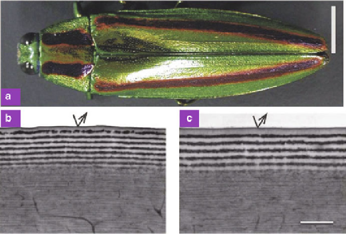
Fig. 8.11
(
a
) Dorsal view of a female Japanese jewel beetle
C. fulgidissima
.(
b
)and(
c
) TEM
images of green and purple sections of the beetle elytra, respectively. Scale bars: (
a
)0.5cm;and
(
b
)and(
c
)1
m (Reproduced from [
50
])
The elytra of some beetles, e.g., the jewel beetles [
21
], display brilliant, metallic
structural colors which are caused by a multilayer in the cuticle. For the buprestid
beetle
Euchroma gigantea
, Durrer and Villinger [
47
] examined its elytra by TEM
and revealed an epicuticle multilayer consisting of five melanin layers with a thick-
ness of 60-80 nm embedded in chitin at a regular distance of 60 nm. This multilayer
was found to be the basis of color production of the elytra. Similar multilayers were
also found in other Buprestidae, e.g., the jewel beetles
Chrysochroa vittata
[
48
,
49
],
Chrysochroa fulgidissima
[
8
,
50
], and
Chrysochroa raja
[
51
].
Figure
8.11
shows the Japanese jewel beetle
C. fulgidissima
[
50
]. The elytra
of this beetle are metallic green marked with longitudinal purple stripes. Because
of the striking iridescence and beautiful luster, this jewel beetle was used as
ornament in ancient Japanese times. TEM characterizations revealed that the
epicuticle of the green and purple areas consists of stacks of 16 and 12 layers,
respectively. The corresponding period for green and purple areas are 160 and 205
nm, respectively. Simulations revealed that both green and purple colors are created
by a multilayer with a surprisingly small gradient refractive-index range of 1.6-1.7.
The multilayer in the cuticle exhibits both strong angle and polarization iridescence,
which is a known optical property for multilayers. The angle dependence can be
understood by the fact that the optical path difference between reflected light at
successive interfaces differs at different incident angles, which leads to iridescence.