Biomedical Engineering Reference
In-Depth Information
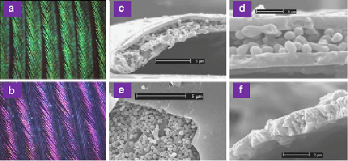
Fig. 8.9
(
a
)and(
b
) Optical microscopic images of iridescent green and purple neck feathers
of domestic pigeons under 100
magnification, respectively. (
c
)-(
f
) SEM images of iridescent
barbules. (
c
) Perspective view of the cross-section of a green barbule. (
d
) Transverse cross-section
of a purple barbule. (
e
) Perspective top view of a green barbule with the top keratin cortex layer
removed (lower left corner). (
f
) Perspective view of the cross-section of a gray barbule. Scale bars:
(
c
)and(
e
)5
m; (
d
)1
m; and (
f
)2
m (Reproduced from [
41
])
shapes of spheres, ellipses, and rods, and also a small amount of randomly dispersed
keratin. The outer surface of the keratin cortex is rather smooth. Green and purple
barbules show different thicknesses. For green barbules, the mean thickness is about
595 nm, while it is about 530 nm for purple barbules. Different from the iridescent
barbules, gray barbules are a single uneven layer composed of randomly dispersed
keratin and melanin granules, leading to gray colors.
The iridescent neck feathers of domestic pigeons display interesting
opposite
iridescence. Namely, green feathers become purple with the observing angle varying
from normal to oblique, while simultaneously purple feathers become green. This
can be clearly seen from the measured reflection spectra for iridescent neck feathers
at different incident angles, shown in Fig.
8.10
. Under normal incidence, iridescent
green feathers display a series of harmonic reflection peaks, positioned at about
415, 530, and 730 nm, corresponding to violet, green, and red colors, respectively.
With increasing incident angle, all reflection peaks show a blue shift to a shorter
wavelength. The violet reflection peak at normal incidence shifts to UV, the green
reflection peak to blue, and the red reflection peak to orange. These harmonic
reflection peaks can be easily understood by thin-film interference. Although there
are two cortex layers, only the dorsal cortex layer gives rise to structural coloration.
This is due to the fact that the central layer sandwiched by the two cortex layers is
composed of randomly dispersed melanin particles such that light will be absorbed
or randomly scattered by this layer. It can also be confirmed by the fact that no
colors can be seen if the dorsal cortex layer is removed. Therefore, the central layer