Biomedical Engineering Reference
In-Depth Information
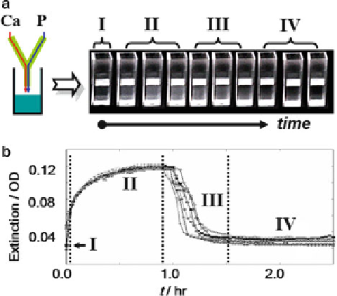
Fig. 7.7
The evolution of calcium phosphate solution. After the mixing of a calcium solution
with a phosphate solution (with final concentration: 4 mM CaCl
2
,6mMK
2
HPO
4
, 150 mM
NaCl, pH 7.40, i.e., the saturation indices (log (
IAP
)-log (
K
sp
), where
IAP
is the ionic activity
product of precipitation;
K
sp
is the solubility product constant) of initial solution were 0.25
(for ACP, Ca
9
(PO
4
)
6
), 25.8 (for HAP), 23.1 (after the precipitation of ACP), the solution
became turbid gradually. In about 1 h, the suspension suddenly became clear, accompanied with
visible sedimentations formed simultaneously (see (
a
)). The entire mineralization process can be
monitored by the extinction curves (the optical difference of 405 nm and 550 nm). (
a
) Photographs
of the suspensions. The solution became turbid, then clear again accompanied with the sediment of
minerals. (
b
) The extinction curves of calcium phosphate solutions. Reproduced with permission
from Ref. [
63
] © The Royal Society of Chemistry 2010
election diffraction (SAED) patterns of diffusive rings indicate that the spheres
are amorphous (Fig.
7.8
a). The initially round-shaped ACP particles indicate the
occurrence of fluid-like structure at the beginning. The sintering of the submicro-
sized ACP droplets in solutions leads to the further aggregation of these spheres
(Fig.
7.8
a). The ACP aggregate partially merged as showed in Fig.
7.8
d. After the
formation of ACP, the particles remained in amorphous state for about 1 h before
the transformation took place.
Concerning the transformation of ACP spheres, at first, the boundary of ACP
spheres became polygonal-like (Figs.
7.8
b,e and
7.9
a; 67 min). Some condensed
rings or dots appear in the SAED patterns (Fig.
7.8
b), indicating the formation
of crystalline materials. The bright dots in the dark-field TEM images (DF-TEM)
(Fig.
7.9
b) indicate the occurrence of crystallized minerals. The high-resolution
TEM images (HR-TEM) (Fig.
7.9
c) directly show that the crystallization happens
at the surface of the ACP spheres, while the main portion of the sphere remains
amorphous (i.e., fast Furrier transform (FFT) patterns of selected region in Fig.
7.9
c,
region 3).