Biomedical Engineering Reference
In-Depth Information
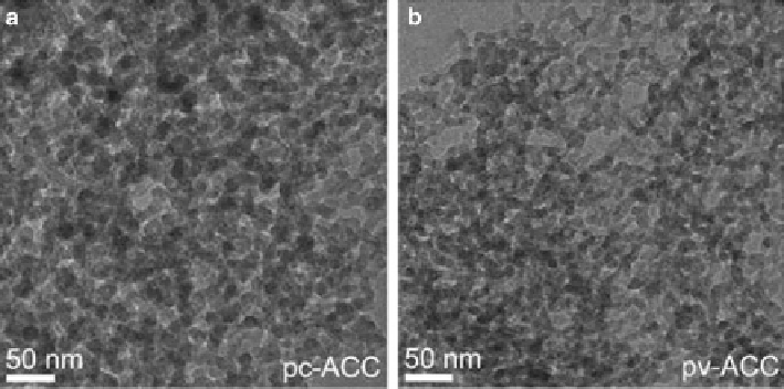
Fig. 6.9
TEM images of pc-ACC and pv-ACC. (Reproduced from [
80
], Copyright © 2010, Wiley)
clearly differs from the crystallization within the droplets. The crystallization was
carried out homogenously by using the Kitano method. These results show that the
ultrasonic trap may be a powerful tool for a real-time analysis of nucleation, crystal
growth, and phase separation processes.
Moreover, a fresh picture of the crystallization of CaCO
3
is emerging, which
involves transformations of clusters to ACC and eventually to crystalline phases
[
78
,
79
]. Hedin and co-workers studied proto-crystalline features of two amorphous
intermediates, ACCI and ACCII, and discussed their relation for crystallization of
CaCO
3
(Fig.
6.9
)[
80
]. They rationalized the identification of ACCI with proto-
calcite ACC and ACCII with proto-vaterite ACC, respectively. These ACCs were
obtained from metastable solutions of CaCO
3
at different pH values by destabiliza-
tion in excess ethanol. Interestingly, they found that structural water in proto-vaterite
ACC and proto-calcite ACC as an explanation of the low coordination numbers for
calcium and deviations from certain symmetries. Therefore, proto-crystalline order
is intrinsic to these ACCs and only one factor influencing crystallization. Additive-
free proto-crystalline ACCs play key roles in the further analyses of the processes
involved in crystallization of calcium carbonate.
6.3.2
Single Crystals Formed via an Amorphous
Precursor Phase
As well-known, calcium carbonate often shows complex single-crystalline struc-
tures, such as the three-dimensionally sculpted conformations of the calcite skeletal
plates of coccoliths and echinoderms [
81
]. Meldrum and co-workers [
82
,
83
]
reported the control of calcium carbonate morphologies and demonstrates that