Biology Reference
In-Depth Information
nonparenchymal cells of the liver, were imaged interacting with the
two-color cancer cells. CFP-expressing host CAFs were predominantly
observed in the TME models developed in the CFP-nude mouse
(
Fig. 10.3
).
22
4. STROMA CELLS ARE REQUIRED FOR CANCER
METASTASIS
After splenic injection of colon cancer cells, splenocytes co-trafficked
with the tumor cells to the liver and facilitated metastatic colony formation.
Extensive clasmocytosis (destruction of the cytoplasm) of the cancer cells
occurred within 6 h after portal vein (PV) injection, and essentially all
the cancer cells died. In contrast, splenic injection of these tumor cells
resulted in the formation of liver and distant metastasis. GFP spleen cells
were found in the liver metastases, which resulted from intrasplenic injec-
tion of the cancer cells in transgenic nude mice ubiquitously expressing
GFP. When GFP spleen cells and the RFP cancer cells were coinjected
Dual color MMT cancer cells
Non
parenchymal cells
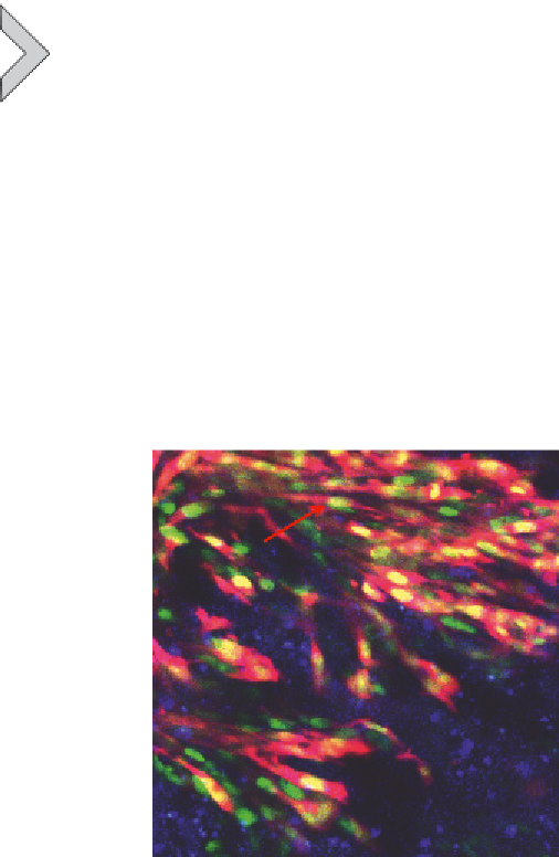
Figure 10.3 MMT cells with GFP in the cytoplasm and RFP in the nucleus, growing in the
liver of a CFP-nude mouse. Dual-color MMT cells formed tumors in the liver of a CFP
mouse 28 days after splenic injection. Hepatocytes, non-parenchymal liver cells (yellow
arrows), and dual-color MMT cancer cells (red arrows) were visualized simultaneously.
The image was taken with an FV1000 confocal microscope. Bar: 50
m.
22
m


Search WWH ::

Custom Search