Biology Reference
In-Depth Information
signal. Cooling the camera reduces stray noise in the electronics and im-
proves the SNR. A secondary image of the tissue or anatomy of interest al-
lows the 2D fluorescent signal to be localized within the animal. One
obvious limitation of 2D imaging is its inability to discriminate signals from
shallow and deep tissue. Fluorescence molecular tomography (FMT) is a de-
veloping technique that offers greater anatomical localization, spatial reso-
lution, and quantitation
56
(
Fig. 9.1
). In FMT, the emitted fluorescence is
measured from a matrix of multiple excitation sources and is normalized
by the
excitation signal
transmitted through the
tissue. Further
100
Preinjection
3h
5h
A
C
Fluorescence (time: 260 min)
(
m
M)
2.5
1.2
GB137
65
1
2
30
0.8
nM
1.5
Preinjection
5h
24h
300
0.6
1
Pro680
200
0.4
0.5
0.2
100
nM
0
0
5h
24h
225
Preinjection
0.5
1
1.5
2
2.5
(cm)
B
GB138
150
1
75
nM
5h
Preinjection
24h
150
0.5
90
Pro750
0
0
50
100
150
200
250
Time (min)
30
nM
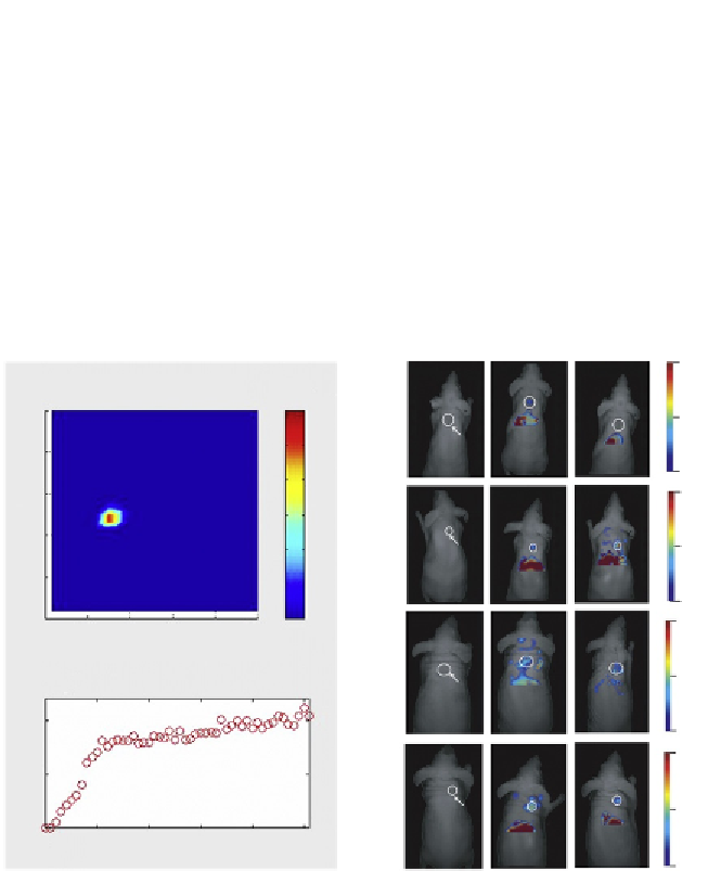
Figure 9.1 Fluorescence-mediated tomography (FMT) in imaging of near-infrared
probe activation in vitro and in vivo: (A) FMT reconstruction image of an NIR fluorescence
source, a 2.5-mm tube filled with a mixture of PGC-Cy5.5 and a model protease (pan-
creatic trypsin) resulting in liberation of fluorescent Cy5.5 dye (1 mM); (B) Time course of
the reaction measured by time-lapse FMT, demonstrating the reaction kinetics resulting
in fluorescence signal shown in (A) (courtesy of Dr. Vasilis Ntziachristos (Technical Uni-
versity of Munich, Germany)); and (C) FMT imaging of the time-dependent accumulation
of small molecular weight and macromolecular probes in a mouse xenograft model of
human cancer. Shown are FMT-reconstructed fluorescent signals in subcutaneous ad-
enocarcinomas of GB137 (quenched activity-based probe); activity-based probe GB138
and macromolecular probes ProSense 680 and ProSense 750. Tumors are shown using
circular regions of interest. Adapted from Ref.
57
.
Search WWH ::

Custom Search