Biomedical Engineering Reference
In-Depth Information
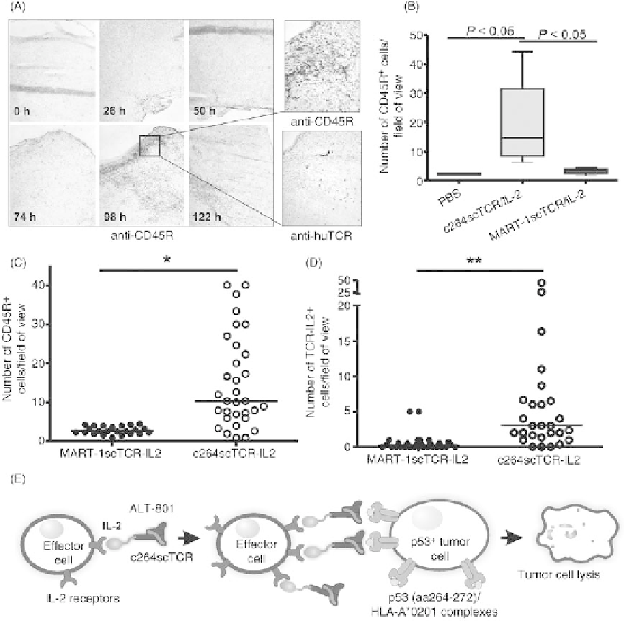
FIGURE 31.4
ALT-801-specific trafficking of immune cells to the tumor site. (A) A375 tumor
bearing nude mice were treated i.v. with ALT-801 (1.6 mg/kg) at 0, 24, 48, 72, and 120 h. At the
indicated times, tumors were collected and cryosections were stained with anti-CD45R and
antihuman TCR mAb. Shown on the left, increasing amounts CD45R
þ
cells (dark stained cells)
were observed in the A375 tumors as treatment with ALT-801 continued for 3-5 days. Tumor
cryosections from the 98 h sample also showed positive staining with the antihuman TCR antibody
indicating cells bearing the ALT-801 fusion protein (see higher magnification on right). (B) Tumor
samples were obtained 24 h post-treatment from A375 tumor-bearing nude mice injected daily for
4 days with ALT-801 (0.5 mg/kg), MART-1scTCR/IL-2 (0.5 mg/kg), or PBS. Tumor sections stained
with anti-CD45R mAb and the positive cells were counted. The median number of CD45R
þ
cells per
field of view is presented in the graph. (C and D) Tumor samples were obtained 24 h post-treatment
from A375 tumor-bearing nude mice injected with ALT-801- or MART-1scTCR/IL-2-coated
splenocytes. Tumor sections stained with anti-CD45R (C) or anti-huTCR mAb (D). Symbols
represent number of positively staining cells per field of view and bars represent median value.
(E) Model of ALT-801 mechanism of action in binding to and activating IL-2 receptor-bearing
immune effector cells and subsequently localizing these cells in the tumor site via pMHC specific
interaction with tumor cells. Source: (A-D) are reproduced with the publisher's permission from
Reference [23].
Search WWH ::

Custom Search