Biomedical Engineering Reference
In-Depth Information
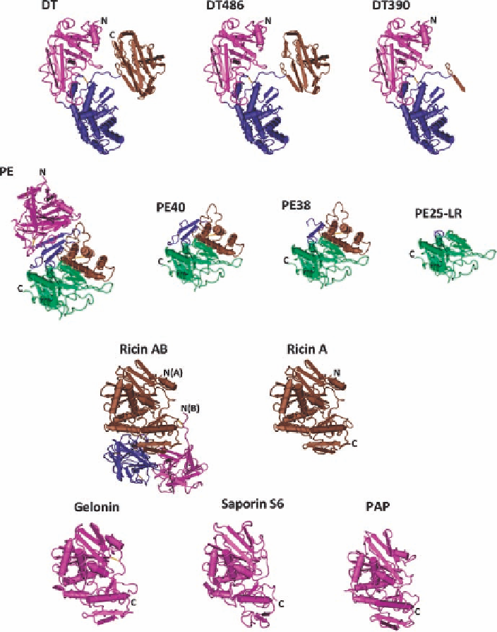
FIGURE 18.1
Backbone diagram of X-ray crystal structures of toxins used in immunotoxin
preparation. N and C indicate N and C termini of proteins, respectively. PAP: pokeweed antiviral
protein. Diphtheria toxin: catalytic domain (magenta), translocation domain (blue), binding domain
(brown), DT (residues 1-535), DT486 (residues 1-486), DT390 (residues 1-390). Pseudomonas
exotoxin: domain 1a (residues 1-252; magenta), domain 1b (residues 365-404; blue), domain II
(residues 253-364; brown), domain III (405-613; green), PE (residues 1-613), PE40 (residues
253-613), PE38 (residues 253-364 and residues 381-613), and PE25-LR (residues 274-284 and
residues 395-613). Ricin: chain A (brown) and chain B (magenta and blue). Ricin, gelonin, saporin,
and PAP have a similar structure.
distinct structural regions: domains Ia (residues 1-252) and Ib
(residues 365-404), domain II (residues 253-364) and
domain III (residues 405-613) [47]. The full-length PE
binds the
a
2
-macroglobulin receptor/low-density lipoprotein
receptor-related protein 1 (LRP1) or LRP1b on the surface
of mammalian cells [48,49]. The PE and receptor complex
is
internalized PE in endocytic vesicles undergoes furin cleav-
age between Arg-279 and Gly-280, a pH-dependent confor-
mation change followed by reduction of the disulfide bond
between Cys-265 and Cys-287. This creates a 28-kDa N-ter-
minal fragment and a 37-kDa C-terminal fragment. The
37 kDa fragment is further routed to the endoplasmic reticu-
lum. In the ER, the 37 kDa fragment is translocated to the
internalized by receptor-mediated endocytosis. The
Search WWH ::

Custom Search