Biomedical Engineering Reference
In-Depth Information
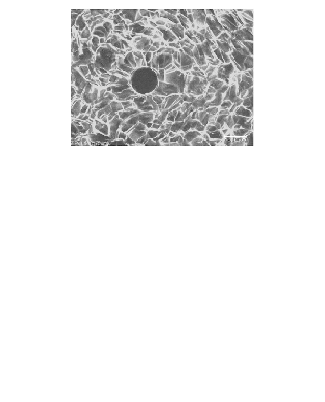
FIGURE 3.2
Confocal microscope image of a blank dual-mode chitosan scaffold.
tissue imaging because it dispenses with the use of fl uorophores and allows the use of near-IR light,
its resolution is lower than that achieved in traditional CM.
130
A new imaging modality, multiphoton confocal microscopy (MCM) has been developed based
on the fact that when photons of certain energy are fi red almost simultaneously to a target mol-
ecule, they can cause an excitation similar to the one produced by the absorption of a single photon
of higher energy. If the excited molecule is fl uorescent, it can emit a single photon of fl uorescence
as if it were excited by a single higher energy photon. This characteristic makes the use of longer
wavelength excitation lights possible (e.g., near-IR), leading to reduced light scattering in tissues
and improved penetration depths, with reduced photobleaching and photodamage. This setup also
simplifi es multicolor imaging by allowing excitation of different fl uorophores with the same laser,
avoiding chromatic aberrations and providing a broad uninterrupted emission collection band-
width. Furthermore, the use of near-IR light favors cell viability. The principles and applications
of this technique are reviewed elsewhere.
131
Again, the limitation of MCM is its low specimen
penetration depth.
3.4.2 M
ICROCOMPUTED
T
OMOGRAPHY
Microcomputed tomography (µ-CT) is an analog of x-ray CT scanner in the medical fi eld. Its
increasing popularity comes from its ability to provide precise quantitative and qualitative infor-
mation on the 3-D morphology of specimens in great detail without resorting to physical section-
ing. Moreover, due to the nondestructive character of the technique and the absence of processing
requirements, the samples can be subjected to other tests eliminating the problem of sample scar-
city. In this imaging technique, the source of contrast is the attenuation of x-rays by high-density
materials. Consequently it has been used in the biological fi eld preferentially for bone tissue
analysis. In µ-CT scanning, the specimen is irradiated from the edges with x-rays in order to
achieve a series of 2-D slices. As the radiation crosses each slice, the x-rays are attenuated, and
the sprouting x-rays with reduced intensities are captured by a detector. The x-ray paths and the
attenuation coeffi cients are derived from the detector measurements. Then, a 2-D pixel map is cre-
ated from these computations, and each pixel is designated by a threshold value that corresponds