Biomedical Engineering Reference
In-Depth Information
10
22
5
SIMS
RBS
4
3
10
21
2
1
10
20
0
0
100
200
300
400
Depth (nm)
FIGURE 19.10
Depth profi le of defects and hydrogen ion concentration in hydrogen-implanted silicon wafer
obtained from RBS and SIMS. (From Liu, X.Y. et al.,
Biomaterials
, 25, 5575, 2004. With permission.)
(a)
(b)
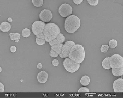
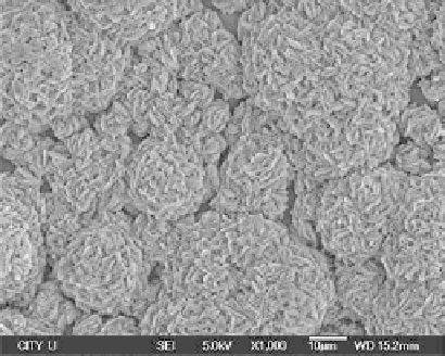
FIGURE 19.11
Surface views of hydrogen-implanted silicon wafer soaked in SBF for (a) 14 days and
(b) 28 days. (From Liu, X.Y. et al.,
Biomaterials
, 25, 5575, 2004. With permission.)
After immersion in the SBFs for 14 days, some single and clustered ball-shaped particles are
observed on the surface of the hydrogen-implanted silicon surface (Figure 19.11a). The surface
of the silicon wafer is, however, not covered completely. After an immersion time of 28 days, the
surface is entirely covered by the newly formed layer (Figure 19.11b). The results obtained from
x-ray diffraction (XRD) (Figure 19.12) and fourier transform infrared (FTIR) (Figure 19.13) show
that carbonate-containing hydroxyapatite (HA) (bone-like apatite) is formed on the surface of the
hydrogen-implanted silicon wafer soaked in SBF, and good bioactivity on the hydrogen-implanted
silicon wafer can be inferred.