Biomedical Engineering Reference
In-Depth Information
determination of the morphology is rather diffi cult since the resolution of phase contrast micro-
scopes is in general too low for determination of platelet morphology. Therefore either fl uorescence
microscopy or scanning electron microscopy is preferred. For fl uorescence microscopy, the platelets
are labeled with a fl uorescent dye, which can be a nonspecifi c viability dye (like Calcein-AM or Cell
Trace dyes). An easy method was discovered by accident.
115
When platelets are fi xed with glutar-
aldehyde, they become fl uorescent, and can be observed by fl uorescence microscopy and counted.
The most used method to observe adhered platelets is scanning electron microscopy. The platelets
are fi xed, and prepared for microscopy by dehydration and sputter coating with gold. It is a straight-
forward method for counting platelets per surface area. Additionally, different morphologies that
occur upon activation of the adhered platelets can be analyzed through this method (Figure 17.8).
50
A disadvantage of such microscopy techniques is that they are relatively time-consuming, very
laborious, and thus expensive. An alternative approach is to determine platelet adhesion by measur-
ing of metabolic parameter like the enzyme lactate dehydrogenase (LDH).
115
The amount of LDH is
a direct measure of the amount of adhered platelets.
17.7.2.2 Platelet Activation
An important parameter upon which the incubation of synthetic surfaces with platelets can be deter-
mined is platelet activation.
5,6,17
Adhered platelets that do not get activated will not contribute to
coagulation. There are three ways to determine platelet activation: (i) platelet morphology, (ii) mem-
brane markers, and (iii) secretion products (Figure 17.17).
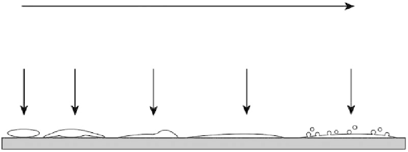
Platelet morphology rapidly changes after initial adhesion and subsequent activation of platelets
on synthetic surfaces.
50
The platelets are a round-shaped just after adhesion and before activation.
Then pseudopods are extended from the central cell body after which the cells fl atten and spread
on the surface until the cell appears completely fl at (like a pancake), covering a large surface.
Upon activation, a number of specifi c markers get exposed on the outer surface of the platelet.
One of these markers is P-selectin (also CD62p), a glycoprotein that has an important function in
interaction with leukocytes. P-selectin can be demonstrated using specifi c antibodies coupled with
fl uorescent labels. Fluorescence microscopy or fl uorescent-assisted cell sorting (FACS) analysis
can be used to determine the amount of exposed P-selectin. Another frequently used marker is the
PAC-1 antibody that binds to the activated integrin α
IIb
β
3
. This integrin immobilizes fi brinogen on
the surface of activated platelets. Use of a fl uorescently labeled specifi c antibody enables the deter-
mination of activation.
Platelet activation
Adhesion
Pseudopods
Spreading
Fully spread
Blebbing
P-selection
ADP/ TX-A2
LDH
PAC-1
Annexin-V
Microparticles
FIGURE 17.17
Schematic representation of platelet adhesion and activation on a synthetic surface. The
markers that can be determined at different stages of platelet activation are given in italics. LDH, lactate
dehydrogenase; PAC-1, antibody specifi c for activated integrin α
IIb
β
3
; ADP, adenosine diphosphate; TX-A2,
thromboxane-A2.