Biology Reference
In-Depth Information
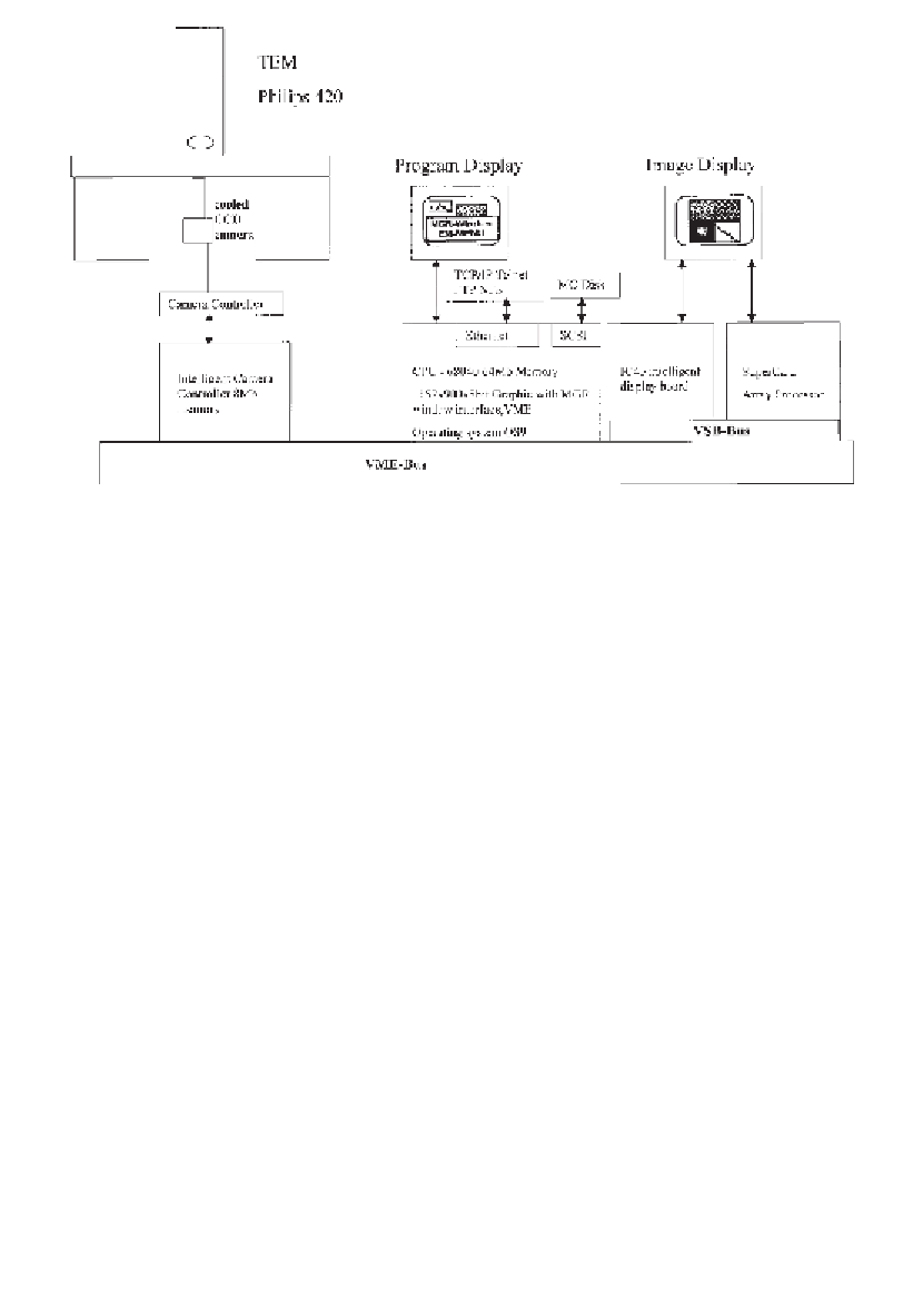
Fig. 4. Diagram to show the TVIPS system used for digital image capture by cooled
CCD. The system allows efficient low-dose imaging and on-line image processing.
7.
In the gap between the two halves of mica, pipet 8
L of ultrapure water. This
forms a “wedge” of water between the mica halves and keeps the adsorbed fibrils
hydrated (
Fig. 5
). (
See
Note 6
.)
µ
8.
Attach the stage to its transfer rod and then plunge it into nitrogen slush to freeze
the sample. After 30 s remove the stage from the nitrogen slush, remove the
spacer from the wedge and then unclamp the Mica Ice Wedge. Reattach it to the
stage by clamping only the lower piece of mica (this allows the top piece of mica
to be removed to facilitate the freeze fracture).
9.
Via the airlock, transfer the stage with the mica ice wedge attached to it into the
CFE-50C, using the transfer rod, for freeze fracturing/etching and rotary shad-
owing. The chamber of the CFE-50C should be at ~2
10
-7
mbar and the stage
×
holder precooled to around -189
°
C (
Fig. 6
).
3.2.2. Fracturing and Etching
1.
The specimen stage is located on a revolving stage holder in the Cressington CFE-
50C. The stage is cooled with liquid nitrogen to keep the temperature at ~ -189
°
C.
2.
Rotate the stage so that the thickest part of the mica ice wedge is facing the liquid
nitrogen-cooled microtome of the Cressington. Lower the microtome blade so
that it will just pass under the top piece of mica in the wedge.
3.
Swing the microtome arm gently toward the sample so that the blade passes under the
top piece of mica. This causes the mica to be lifted free and clear of the ice wedge,
thereby producing the fracture that exposes the surface of the sample (
Fig. 7
).
Search WWH ::

Custom Search