Biology Reference
In-Depth Information
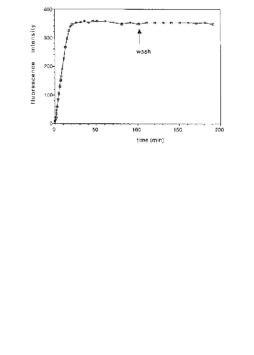
Fig. 3. Kinetics of formation of supported planar bilayers by fusion of DMPG/
DMPC vesicles containing integrin
3. Integrin was fluorescently labeled with
FITC prior to incorporation into DMPG/DMPC vesicles. The cell was filled with
Buffer B and vesicle suspension (about 50 nmol of lipid) was injected into the measur-
ing cell containing vesicle fusion buffer to let the vesicles fuse onto a quartz slide.
Fusion kinetics were followed by measuring the increase of fluorescence intensity by
TIRFM. After 100 min the cell was washed with buffer to remove excess of vesicles.
No change in fluorescence intensity was observed after the washing step (arrow).
α
IIb
β
close agreement with earlier data obtained earlier
(18
,
19)
and clearly indicate
the intactness of the membrane with free mobility of the fluorescently labeled
lipid in both leaflets of the supported bilayer.
The lateral diffusion coefficient of the FITC-labeled
3 was found to be
only six times smaller than that of the lipid. The value of 0.70
α
IIb
β
10
-8
cm
2
/s
corresponds well with values found for other monodispersed membrane-span-
ning proteins at comparable surface concentrations
(20)
. FRAP also revealed
that about 50% of the integrin population was mobile (
Fig. 4
) and the immobil-
ity of the remaining population is consistent with integrins oriented with their
large extracellular part towards the quartz support of the bilayer, allowing
attachment of these domains to the support. In the mobile population, the small
size of the cytoplasmic domains prevented the integrin from sticking to the
support
(21)
.
Figure 2C
depicts a schematic drawing of the orientation of the
integrins in supported planar bilayers.
×
Search WWH ::

Custom Search