Biology Reference
In-Depth Information
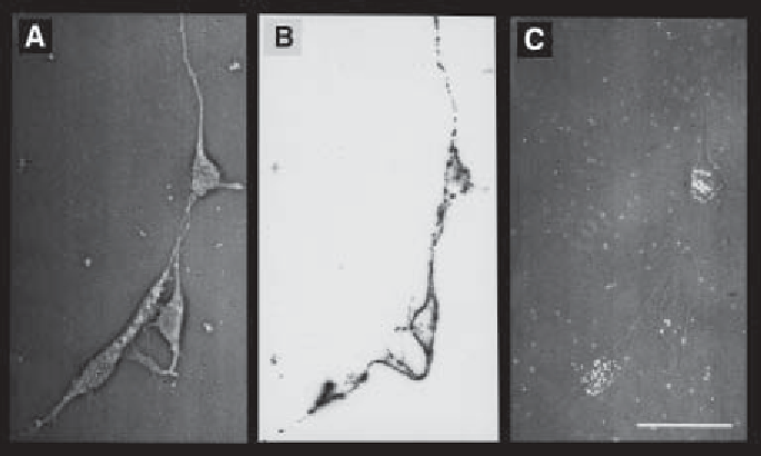
Fig. 2. Effects of laminin-1 on cultured E5 retinal cells. Dissociated cells were
cultured on polyornithine/laminin-1 in the presence of 0.5
Ci
/mL
[
3
H]thymidine.
After 20 h in culture, cells were immunolabeled with G4 mAb (specific for neurons)
and subjected to autoradiography. Cultured cells were finally observed under phase-
contrast
(A)
, fluorescence
(B)
, and bright-field
(C)
microscopy. Observe the presence
of G4-positive, [
3
H]thymidine-positive neurons. These cells were still in process of
cell division at the moment of plating and have acquired neuronal properties during
the time in culture (i.e., in vitro differentiated neurons). From
(8)
(with permission).
µ
8.
Cultures are then dehydrated by passing the coverslips through 50%-, 70%-, 96%-
and absolute ethanol. The coverslips are stuck to a glass slide with DePex and
then covered with NTB-2 photographic emulsion and exposed for 2-3 d in the
darkness. 100
L photographic emulsion should be laid onto the coverslip sur-
face containing the cells and quickly removed to leave only a thin film covering
the cells.
µ
9.
Develop the photographic reaction for 2 min using D19 developer diluted 1:1
in water. Wash once in water, fix in 25% sodium thiosulfate for 5 min and wash
extensively with water. Finally, mount coverslips with Ca
2+
-, Mg
2+
-free PBS/
Glycerol and observe under a fluorescence microscope. Cells double labeled for
G4 and [
3
H]methyl thymidine are considered to be in vitro differentiated neurons
(
see
Fig. 2
).
3.3. Differentiative Bioassay Using Soluble ECM Molecules
Unlike the protocol described previously, which lacks appropriate controls,
the protocol described here uses fibronectin as a control substratum, the ECM
molecule to be tested is added to the medium subsequently. This protocol is

Search WWH ::

Custom Search