Biology Reference
In-Depth Information
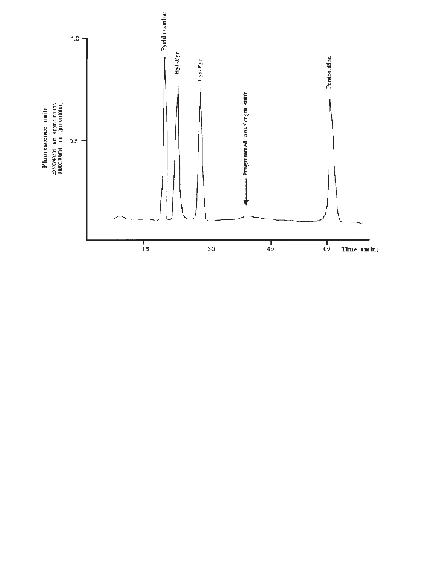
Fig. 3. Relative elution positions of the standards, pyridoxamine, hydroxylysyl-
pyridinoline (Hyl-Pyr); lysylpyridinoline (Hyl-Pyr) and the glycation crosslink
pentosidine on a Hypercarb S reversed phase HPLC column using fluorescence detection.
6.
After an 8 min isocratic period in water, a 0-12% THF linear gradient in water is
applied over 60 min at a flow rate of 1 mL/min (0.2%/mL/min). Hydroxylysyl
and lysyl-pyridinoline elute at approx 26 and 29 min, respectively, and the
glycation crosslink pentosidine elutes at 62 min.
7.
The pyridinium crosslinks are detected by means of their natural fluorescence at
405 nm emission after excitation at 295 nm. Pentosidine is also naturally fluores-
cent but at 385 nm emission after excitation at 335 nm. We program a wave-
length shift into our Perkin-Elmer LS-5 fluorimeter (Bucks, UK) to take place
after the pyridinolines have eluted. A typical HPLC elution profile of pyridinoline
and pentosidine standards is shown in
Fig. 3
.
8.
In this laboratory, data are collected during the analytical run using a computing
integrator and stored to disk at the end of the analysis.
9.
The area under each peak of interest is calculated as a proportion of that derived
from known concentrations of standards prepared within this laboratory or pur-
chased commercially. Where possible, the concentration of the standards should
be confirmed by amino acid analysis.
10.
Column integrity and fluorimeter efficiency is confirmed by regularly running a
standard mixture (every 8-10 samples) and calculating the fluorescence yield per
pmol of each fluorophore.
Search WWH ::

Custom Search