Biology Reference
In-Depth Information
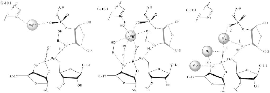
Figure 2.6 Schematic view of the coordination sites in the HHR active site. Left: The coordination pattern of Mg
2þ
in the
“
C-site
”
coordinated
to G10.1:N7 and A9:O2p. Middle: The coordination pattern of Mg
2þ
in the
bridging A9:O2p and C1.1:O2p of the scissile phosphate.
Right: Coordination sites for Na
þ
in the HHR active site found in the RT-Na and dRT-Na simulations. Red numbers next to the coordination sites
are the scores used to calculate the coordination index (see text). M1 involves direct binding to A9:O2p and C.1:O2p and indirect binding to
G10.1:N7 through a water molecule. M2 involves direct binding to C17:O2
0
and C.1:O2p. M3 involves direct binding to C17:O2
0
and is posi-
tioned toward the outside of the active site.
“
B-site
”
Search WWH ::

Custom Search