Biology Reference
In-Depth Information
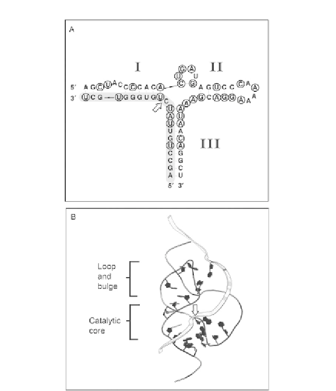
Figure 1.5 Mapping of CLEC2 ribozyme invariant nucleotides on the tertiary structure of
the full-length hammerhead ribozyme. (A) Secondary structure of mCLEC2d hammer-
head ribozyme. Positions conserved in all CLEC2 ribozyme sequences are circled. The
cleavage site is indicated with a white arrow. (B) Positions analogous to invariant CLEC2
ribozyme nucleotides are drawn in black on the string representation of the Schistosome
hammerhead ribozyme tertiary structure (PDB ID: 2GOZ).
41
The substrate strand is rep-
resented as a wide ribbon and the site of bond cleavage is indicated with a white arrow.
ranges from 246 to 789 nt. Nevertheless, the substrate and enzyme segments
pair to form a structurally and catalytically accurate hammerhead ribozyme.
More specifically, CLEC2 ribozymes contain the signature core of 15 invari-
ant nucleotides flanked by three helices with distal rate-enhancing interac-
tions, and they produce two strands by self-cleaving (
Fig. 1.4
). Even though
Search WWH ::

Custom Search