Biomedical Engineering Reference
In-Depth Information
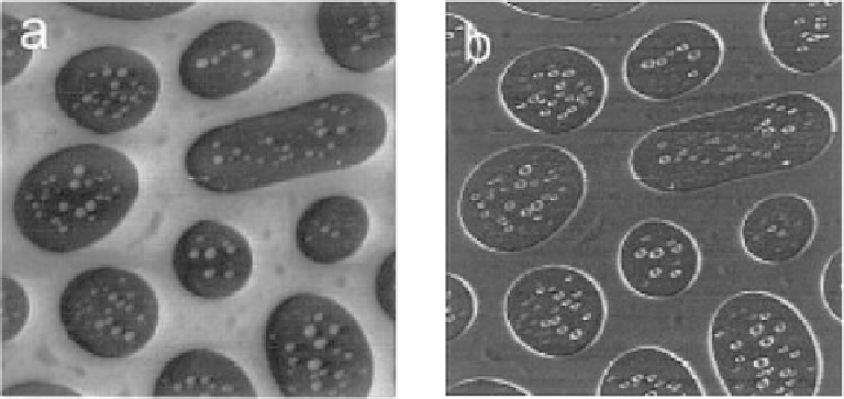
Fig. 3.20. Example of pulsed force mode. The sample is a polystyrene-polymethylmethacrylate
blend. A: topography, B: adhesion, both measured simultaneously. Note the bright borders between
the phases are due to increased tip-sample contact area, and the adhesion image is in agreement with
that measured by force spectroscopy [159]. Reproduced from [206] with permission.
with jumping mode AFM, a major aim of pulsed-force AFM is to obtain adhesion data
[208], but collection of other data points can again lead to sample stiffness data [199]. An
example of the results from pulsed force mode is shown in Figure 3.20.
3.2.4 Magnetic force microscopy
The potential of using AFM to measure magnetic properties was realized quite early in the
history of AFM [105, 209, 210]. Magnetic fields decay quickly with distance, so in order
to measure local properties the probe must be very close to the surface, hence the
applicability of AFM. The most typical experiment carried out is known as magnetic
force microscopy (MFM) [211]. In this mode, the presence and distribution of magnetic
fields is measured directly, by using a magnetic probe. Typically, these consist of standard
silicon cantilevers with a thin magnetic coating. Typical materials used for the coating
include cobalt, cobalt-nickel and cobalt-chromium [212]. The addition of such coatings
can have two detrimental effects on the cantilever: firstly these materials are typically
softer than the underlying silicon, and thus may increase wear rate, and secondly, any
coating added to the end of the tip will increase the radius, and thus decrease the resolution
of the experiment. Typically, magnetic forces are orders of magnitude lower than other
tip-sample forces when in contact, and thus it is useful to measure them with the tip at a
certain distance (of the order of 5-50 nm) from the surface, thus reducing the interference
from short-range forces. This can be carried out in a number of ways [213], some of which
are illustrated in Figure 3.21. These techniques all have some practical advantages and
disadvantages, but are basically variations on a theme. In 'lifting'-type modes, the
topography of the sample is measured first, followed by raising the probe, and scanning
again to collect the magnetic data. One method is to collect a normal topography scan, and
then change the
z
set-point to lift the probe from the surface and collect a 'magnetic image'