Biomedical Engineering Reference
In-Depth Information
30
9 nm
Bacterial spores control
Bacterial spores treated
20
0 nm
10
Bacteria control
Bacteria treated
0
500 nm
0
50
100
Displacement (nm)
150
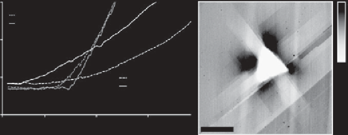
Fig. 3.17. Examples of nanoindentation measurements with the AFM. Left: force-distance curves
measured with the AFM on individual bacteria. Black curves: typical data measured on untreated
and treated
Bacillus
vegetative bacterial cells. Red curves: data measured on
Bacillus
spores.
The data showed that the treatment made the cells softer, but the spores were much harder than
the vegetative cells [178]. Right: AFM image of an indentation made by a dedicated nanoindenter.
The indentation is in a magnesium oxide crystal, and the image shows the indentation (black
triangle) pile-up - material pushed out of hole (white features at triangle corners), and also shows
long-range dislocations in the crystal structure (diagonal discontinuities) [179]. Reproduced with
permission from [180] and kind permission from Dr C. Tromas.
for example individual micro-organisms [169, 183] (see Figure 3.17), living cells
[176, 184] or micro/nanoparticles [185-187]. Some more examples of applications of
nanoindentation are given in Chapter 7.
3.2.3 Mechanical property imaging
Nanoindentation is a very useful technique for mechanical characterization because of the
possibility to collect truly quantitative data on the mechanical resistance of samples. However
it has several drawbacks, including the complicated data analysis, and its relatively slow data
acquisition. The very low rate of data acquisition compared to normal imaging AFMmodes is
a major drawback. For an image with 512
512 data points, a full set of nanoindentation data
would require many hours to collect, leading to problems with thermal drift of the sample. For
this reason 'imaging' type studies with nanoindentation tend to be used only at very low
resolutions (100
100 data points or less). One way to overcome this limitation is to measure
the interaction of the probe with the sample surface while it acquires topographical data, and
use this information to derive mechanical information about the sample surface. This has two
advantages, firstly, data is acquired at a much faster rate, and secondly, the mechanical
information collected may be correlated directly with the measure topography. There are a
number of modes which acquire mechanical information about the sample surface in this way,
and they are described in the following sections.
3.2.3.1 Lateral force microscopy
As described in Section 3.1.1, in contact mode, the vertical deflection of the cantilever,
measured as the difference in signal between the top and the bottom of the split photodiode,