Biomedical Engineering Reference
In-Depth Information
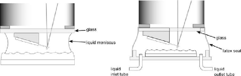
Fig. 2.35. Two approaches to AFM scanning in liquid with a probe-scanning microscope. Scanning in
a droplet of water (left) is simpler, but allows less control of the solution. Scanning in an AFM fluid cell
(right) allows exchange of fluids, and control of fluid temperature, ionic strength, etc.
damage from water from the liquid cell. Due to the specific danger of piezo damage,
AFMs designed for liquid work are usually based on the tip-scanning design, to
minimize the chance of liquid reaching the scanner. AFM liquid cells will typically
have ports to exchange fluids during scanning, and can be of a semi-closed or com-
pletely sealed design. With sealed cells, some flexibility (a rubber membrane or o-ring)
must be included to permit the movement of the piezo, or of the sample. Other typical
additions to liquid cells are electrical connections (for electrochemical AFM), or heat-
ing/cooling circuits (see next section). An alternative to a liquid cell for scanning in
fluid is to simply scan within a droplet of water. An illustration of this technique and of
a closed liquid cell is given in Figure 2.35. In this method, typically applied with probe
scanning microscopes, the bottom of the scanner assembly is covered by a glass
window, to allow the laser to focus on the cantilever, which is mounted below the
glass window. A droplet of liquid sits on the sample surface, and when the scanning
head is lowered into it, the liquid forms a continuous bridge between the glass of the
scanning head, and the sample surface. This technique only works with aqueous
samples, and hydrophilic samples, as the surface tension of the water is required to
keep the droplet in place as the AFM head is lowered. Furthermore, the droplet is prone
to evaporate over a period of time. With these caveats, however, the technique is
surprisingly effective, and greatly reduces the chances of bubbles entering the optical
path, which can cause problems in closed cells. Non-aqueous solvents must be scanned
in the closed liquid cell. More details about procedures for scanning in liquids are given
in sections Chapter 4.
Temperature control
Temperature control of scanning is highly desirable for a wide range of applications in
AFM. In fact, AFM has been performed at a very wide range of temperatures; imaging at
temperatures as low as 5 K [1, 87] and as high as 750
8
C [88]. However, for practical
purposes, with commercial AFMs, in air, a more limited range of temperatures is avail-
able, due to two main effects:
• at low temperatures - condensation on the sample or microscope;
• at high temperatures - destruction of the AFM scanner.