Biomedical Engineering Reference
In-Depth Information
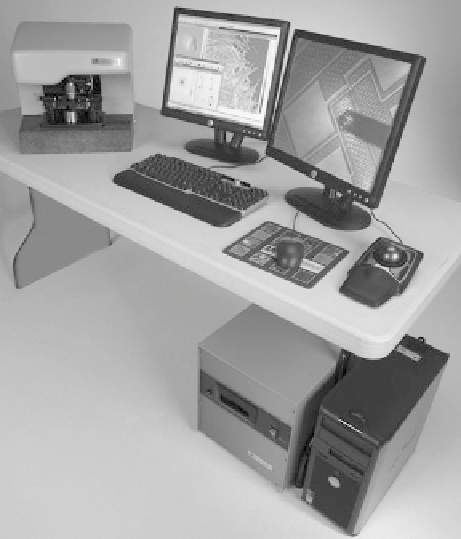
Fig. 2.1. Photo of a desktop AFM illustrating the major components. They are the microscope stage,
computer, electronic controller, computer monitor, and optical microscope monitor. The trackball is
used for moving the sample stage in the
X-Y
axis. Resolution can usually be improved by placing the
microscope stage on a vibration isolation table.
material, the geometry of the device, and the magnitude of the applied voltage. This is
illustrated schematically in Figure 2.2.
Typically, the expansion coefficient for a single piezoelectric device is on the order of
0.1 nm per applied volt. Thus, if the voltage used to excite the piezomaterial is 2 volts,
then the material will expand approximately 0.2 nm, or approximately the diameter of a
single atom. It is the ability to accurately control such tiny movement that makes
piezoelectric materials so useful for AFM. Thus, piezoelectric materials are used for
controlling the motion of the probe as it is scanned across the sample surface. Piezo-
electrics are available in a variety of sizes and shapes, and are generally used in more
complex geometries than depicted in Figure 2.2, so that they can scan the tip in multiple
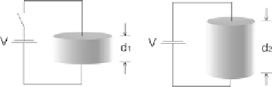
Fig. 2.2. A piezoelectric disk will expand radially (
d
2
> d
1
) when a voltage potential is applied to
the top and bottom electrodes. The disk will change shape such that volume is preserved.