Biomedical Engineering Reference
In-Depth Information
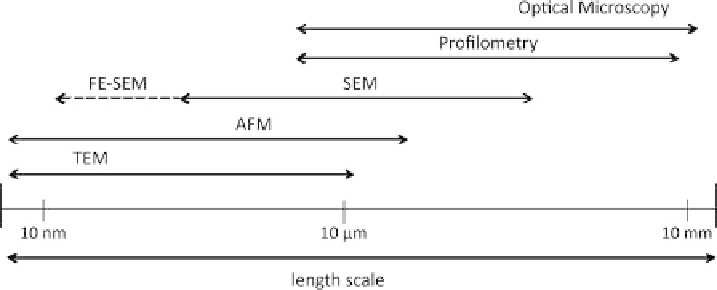
Fig. 1.6. Comparison of length-scales of various microscopes.
not practical to make measurements on areas greater than about 100
m. This is because
the AFM requires mechanically scanning the probe over a surface, and scanning such large
areas would generally mean scanning very slowly. Exceptions to this include parallel
AFM that measure small areas but with many probes to build up a large dataset, or 'fast-
scanning' AFMs, which are discussed in Chapter 2.
When compared to a profiler, the AFM has a greater
X-Y
resolution because in the AFM
the probe is sharper. The fine control of probe-surface forces enabled by this feedback
mechanism enables the use of lower loading forces, which allows the use of much sharper
probes, resulting in much higher
X-Y
resolution. The difference in applied force is very
high, while profilometers will typically apply
ca
.10
6
N to the surface, AFMs can image
with 10
9
N or less. Profilers can have high vertical resolutions, as low as 0.5
˚
. However,
much greater bandwidth in the AFM experiments means that practically, the AFM height
resolution is far greater than that of the profilometers. This is because the bandwidth limits
on profilometers mean that to achieve high height resolution scanning must occur very
slowly.
The length-scale of an optical microscope overlaps nicely with an AFM. Thus, an AFM
is often combined with an optical microscope and with this combination it is possible to
have a combined field of view with a dynamic range from mm to nm. In practice, a
simplified optical microscope, known as an inspection scope, is usually used for selecting
the location for AFM scanning. However, a combination of high-resolution optical
microscopes, often with fluorescence microscopy integration, with AFM also has great
advantages, especially in biology. This is discussed further in Chapter 2 and in Section 7.3.
The combination of AFM with other microscopes or instruments is made simple by the
AFM's small size.
The AFM is most often compared with the electron beam techniques such as the
Scanning Electron Microscope (SEM) or Transmission Electron Microscope (TEM). As
may be seen in Figure 1.6, the dimensional range of these techniques is rather similar, with
SEM (usually) having a somewhat lower resolution to AFM, while the ultimate resolution
of TEM is quite similar to that of AFM. Table 1.1 contains a list of some of the major
factors in comparison of AFM with SEM and TEM.
In general, it is easier to learn to use an AFM than an electron microscope because there
is minimal sample preparation required with an AFM, and nearly any sample can be