Biomedical Engineering Reference
In-Depth Information
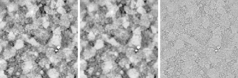
Fig. 5.4. Example of the results of matrix filtering. Left: unfiltered 2
m image of ITO-coated glass.
Middle: result of filtering the image with a 5
5 mean filter; this results in smoothing of the image
and reduction of high-frequency noise and details. Right: result of filtering the image with a 5
5
unicrisp filter. This reduces low-frequency height differences, but greatly enhances edges and high-
frequency features.
There are many other matrix filters that can be used, and are typically available in
AFM processing software. These are more sophisticated versions of the filters
discussed above, some of which, such as the median filter, can reduce noise without
inducing edge-blurring as shown above for the mean filter [352]. Fourier transform-
based filtering relies on transforming an image from real space into frequency space,
removing certain components, and transforming back into an image in real space.
Since Fourier transforms are used more often in AFM as an analysis tool than as a
processing tool, they are discussed further in Section 5.3. In terms of their use as a
filtering tool, Fourier techniques can achieve very similar results to matrix filtering,
i.e. either high- or low-pass filters can be applied. However, Fourier filtering is
somewhat more flexible, and it is possible to remove or to enhance specific com-
ponentsofanimage,asshowninSection5.3.4.
5.1.3 Rotation, cropping, and scaling
Rotation of images is a commonly available procedure in AFM analysis. This is necessary
if it is required that features in an image line up with the scan axis. This can be required for
the analysis of technical samples, for example for specific software analysis routines. In
addition, occasionally it is necessary to scan a particular part of a sample before and after
treatment. In this case, it can be useful to align the images before and after treatment.
A common treatment applied to AFM images is cropping, which could be used to
remove unwanted features from the edges of the scan, or to isolate a particular part of the
image for further analysis. For example, the roughness of different regions in a sample
could be analysed by this technique. Pixelation caused by zooming into small regions of an
image can make the image difficult to interpret. For display purposes, additional pixels can
be added and a resampling algorithm function used to 'smooth' the image. However, this
does not add any additional data to the image, and resampled images should not be used
for further analysis.
Scaling in this context means changing the scale of an image. This is an operation that
will rarely need to be used, as the calibration of the AFM instrument should be correct