Biomedical Engineering Reference
In-Depth Information
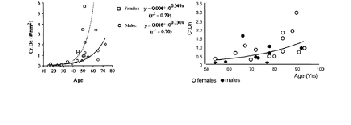
Fig. 5 Microcrack density in (left) cortical and (right) cancellous bone shows an exponential
increase with age in both males and females. Reprinted with permission from Elsevier and John
Wiley & Sons [
1
,
31
]
bone specimens from older human donors with an average age of 70.5 and
77.9 years for men and women, respectively [
6
]. It is unclear whether the elevated
microdamage in the old females in this study reflects greater relative accumulation
in the early post-menopausal years (when there is elevated bone turnover) or in the
later years after post-menopausal bone loss has occurred.
In contrast to linear microcracks, the identification and measurement of diffuse
damage is a relatively new area and its relationship to age, gender, and bone
remodeling is still unclear. For example, Vashishth et al. found that diffuse damage
in cancellous bone is compartmentalized primarily near trabecular surfaces, which
is readily accessible for repair by surface based remodeling [
15
]. There was no
age-related trend in male or female groups but more diffuse damage was present in
men than in women (age range = 23-96 years) [
15
]. Since the women in this
study were post-menopausal age, typically associated with high bone turnover
rates [
43
], the existence of more diffuse damage in males than females could be
due to differences in bone turnover. In contrast to Vashishth et al. [
15
], Arlot et al.
did not find gender based differences, but detected an age-related accumulation of
diffuse damage in trabecular bone (age range = 54-93 years) [
31
]. This investi-
gation included a larger proportion of older donors, and the results may be
indicative of changes in bone due to senile osteoporosis.
Studies conducted by the author's group and others have shown the mechanical
effects of diffuse damage on bone fracture (see next section for details). However,
the biological consequences of diffuse damage including damage initiated
remodeling are largely unknown. To date, only a single study by Bentolila et al.
[
30
] has examined such a relation between diffuse damage and bone resorption.
Unlike linear microcracks, Bentolila et al. found only a statistically non-significant
trend between diffuse damage and bone resorption in rats. Furthermore, unlike
microcrack initiated osteocyte apoptosis [
34
], no mechanism for diffuse damage
initiated bone resorption has been reported. Because diffuse damage in aging
human cortical bone decreases with age [
8
], it is likely that diffuse damage triggers
a biological response for its repair and/or reduction. More studies are needed to
examine bone's in vivo response to diffuse damage.
Search WWH ::

Custom Search