Biomedical Engineering Reference
In-Depth Information
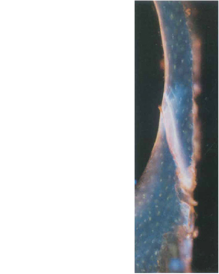
Fig. 2 A microcrack (light
blue and white) was labeled
with calcein blue after the
first 75% of fatigue testing
followed by xylenol orange
after the last 25% of the test.
Reprinted with permission
from John Wiley & Sons [
20
]
Following the completion of the fatigue testing, specimens are incubated in
ethanol for dehydration, embedded in methyl methacrylate, and ground to a
thickness of 100 lm. Results obtained during the development of this protocol
showed microcrack growth during the duration of fatigue testing [
20
], and
illustrated the use of dye combinations to capture microdamage development and
accumulation [
23
].
The imaging techniques for microdamage characterization discussed above
are limited to two-dimensional histological sectioning although microdamage
extends in three dimensions. Recently, three-dimensional methods for microdamage
Search WWH ::

Custom Search