Biomedical Engineering Reference
In-Depth Information
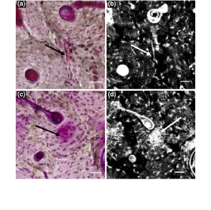
Fig. 1 Microdamage forms as two different morphologies. An image of a linear microcrack (top)
is shown under a bright-field microscopy, and b confocal microscopy. An image of diffuse
damage (bottom) is shown under c bright-field microscopy, and d confocal microscopy. Scale
bars = 50 lm. Reprinted with permission from Elsevier [
8
]
a repair process of intracortical remodeling [
3
-
5
]. However, there is an age-related
accumulation of microdamage in bones [
1
,
6
], due either to deterioration of the
repair mechanism with age [
4
,
7
] or to matrix changes that reduce the damage
resistance of bone.
Microdamage can take two distinct forms [
1
,
6
,
8
]. Two damage morphologies,
linear microcracks and diffuse damage (Fig.
1
), result from different types of
applied loading [
9
-
11
]. Linear microcracks form primarily due to compressive
loading and appear as sharply defined cracks [
10
,
12
,
13
]. They are primarily
found in the interstitial regions of bone where they follow the lamellar interface
and stop at the cement lines of osteons [
8
,
11
,
14
]. On the other hand, diffuse
damage results from tensile loads [
9
]. It has the appearance of a spread mesh of
submicroscopic cracks [
15
]. Diffuse damage is closely associated with osteonal
regions in bone, and it does not follow the lamellar boundaries [
8
].
Search WWH ::

Custom Search