Biomedical Engineering Reference
In-Depth Information
Fig. 2 Lamella can be
considered as a sheet of long
fiber reinforced composite
Cortical bone
Osteon
Osteon
Lamella
Interstitial
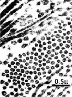
Fig. 3 Collagen fibril
distributions in a lamella
of bone
have a preferred but shifting orientation (Fig.
3
). With reflective light microscopy,
lamellae in osteons appear as white bands of varying thickness separated by a thin
dark layer, which results from the difference in the orientation of collagen fibrils
between neighboring lamella. Two general architectures of lamellae are postu-
lated: (1) ''orthogonal plywood'' with alternating orthogonal orientations of fibrils,
and (2) ''twisted plywood'' with continually changing orientation of fibrils in
which the pattern repeats itself through 180 cycles [
37
]. TEM and SEM obser-
vations also show that parallel fibrils may rotate at a plywood angle of *30
through several successive sub-layers of varying thickness in a lamella [
38
,
39
].
The fibrils may intermingle across lamellae, but there is a distinct and preferred
orientation for any given layer [
5
].
2.3 Ultrastructure of Cortical Bone
The nano-scale structure (often called ultrastructure) and the interactions between
mineral, collagen and water in bone are still poorly understood. For example, some
investigators argue that most mineral crystals reside in the intrafibrillar spaces
(e.g., gap regions) during mineralization of collagen fibrils [
40
,
41
]. However,
some studies indicate that only limited portion of the mineral phase is in the
intrafibrillar space, whereas a large percentage of mineral crystals are deposited
outside of collagen fibrils [
42
,
43
].


Search WWH ::

Custom Search